Anatomy of Human Ear-Process of Hearing and Functions
 written by Sidra Batool
The ear is one of the most important organs in the human body. It is responsible for detecting and transmitting sound. The human ear, The human ear doesn’t just allow us to hear but also keep our balance. Other mammals have similar ear structures that help them with both hearing and maintaining their equilibrium.
For humans, the sense organs in our ears help us to coordinate our head and eye movements ears are organs that provide two main functions: hearing and balance. These functions depend on specialized receptors called hair cells.
Also Read: What is Anatomy?
Structure Of Human Ear
The human ear has three divisions: the outer, middle, and inner ear;
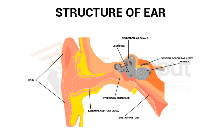
1. Outer Ear
The outer ear consists of the auricle and external auditory canal.
2. Middle Ear
The middle ear begins at the tympanic membrane and ends inside the skull small membranous openings are located in the middle ear. These are called ossicles. and round windows. Three small ossicles (bones) are present between the tympanic membrane and the oval window. They are named according to their shapes. These ossicles are:
1. Malleus (hammer): It adheres to the tympanic membrane and connects the incus.
2. Incus (anvil): The incus connects to the stapes.
3. Stapes: It adheres to the oval window.
An auditory tube or Eustachian tube is present in the middle ear. It extends from the middle ear to the nasopharynx. It equalizes air pressure between the middle ear and the throat.
3. Inner Ear
The inner ear has three components: Vestibules, semicircular canals cochlea. The first two components vestibule and the semicircular canals concerned with equilibrium. There are three semicircular canals. Each canal is arranged in each dimension of space. The third component of the cochlea is involved in hearing.
Process Of Hearing
The following steps are involved in the process of hearing.
1. Sound waves enter the outer ear. It creates pressure waves. These pressure waves reach the tympanic membrane.
2. The pressure waves vibrate the tympanic membrane. The vibrations mow the Malleus on the other side of the membrane.
3. The handle of the malleus articulates with the incus and vibrates it.
4. The vibrating incus moves the stapes back and forth against the oval window.
5. The movements of the oval window set up pressure changes. It vibrates the fluid in the inner ear. These vibrations are transmitted to the basilar membrane. The basilar membrane starts vibrating.
6. Organ of Corti is present on the basilar membrane. They contact the overlying tectorial membrane. This membrane bent. It produces a generator potential. It causes an action potential. This action potential travels along the vestibule-cochlear nerve and reaches the brain for interpretation.
7. The movements of the round window dissipate the vibrations in the cochlear fluid.
Humans can hear low-pitched sounds, below 20 cycles per second. Young children can hear high-pitched sounds up to 20,000 cycles per second. This ability decreases with age. Other vertebrates can hear sounds at much higher frequencies. For example, dogs can easily detect sounds of 40,000 cycles per second. Thus, dogs can hear sounds from a high-pitched dog whistle. A human cannot hear this whistle.
Function Of Ear In Hearing And Balance
The sound waves travel through the ear canal. The tympanic cavity is connected to the tympanic membrane. The stapes push the window in and out of the tympanic cavities. This action is passed on to the organ of the Corti, which has tiny hair cells that translate the sound into an electrical impulse that is transmitted to the brain.
The middle ear air pressure is equalized by the eustachian tube. Body balance is maintained by the vestibular complex.
Leave a Reply