Human Ear Anatomy Diagram Explained
 written by Sidra Batool
Human Ear Anatomy
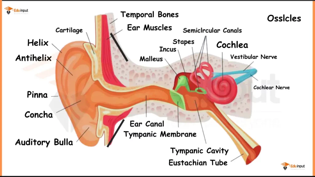
Here are the main parts of Human Ear shown in above diagram:
1. Pinna (Auricle) – In above diagram, the visible, external portion of the ear is Pinna. It is commonly known as earlobe. It is a flexible structure that is composed of elastic cartilage covered by skin. It collects incoming sound waves and channel them towards the inner ear.
2. External Auditory Canal – It is a short, slightly curved tunnel that leads from the pinna inwards. This is lined with fine hairs and ceruminous glands (wax-producing glands). These hairs help trap dust and debris, while the wax acts as a protective barrier against dust particles and insects. Sound waves travel down this canal and reaches to the eardrum.
3. Eardrum (Tympanic Membrane) – This is a thin, tightly stretched membrane that separates the external auditory canal from the middle ear. It acts as a miniature drum, that vibrates in response to incoming sound waves.
4. Ossicles – Now we enter the middle ear. It is a small air-filled cavity. Here chain of three tiny bones are present that are known as the ossicles. These bones are the:
- Malleus (Hammer Shaped) – It is the first bone, that is connected to the eardrum.
- Incus (Anvil-like shape) – The middle bone receives vibrations from the malleus.
- Stapes (Stirrup shaped) – It is the smallest bone in the human body. It transmits the vibrations further into the inner ear.
The ossicles work together to amplify the vibrations received from the eardrum.
5. Cochlea: After Ossicles, you can see the inner ear. This is a complex maze-like structure filled with fluid. The cochlea is the most important part of the inner ear. It is a spiral-shaped cavity that looks like a snail shell.
6. Semicircular Canals are present near the cochlea. It is a set of three fluid-filled loops. These canals are responsible for maintaining our sense of balance by detecting head movements and body orientation.
7. Auditory Nerve – Finally, you can see the auditory nerve in this diagram. It receives the electrical signals generated by the hair cells within the cochlea due to sound vibrations. These signals are then transmitted to the brain, where they are interpreted as sound.
Leave a Reply