What is the Countercurrent Exchange Mechanism?
 written by Sidra Batool
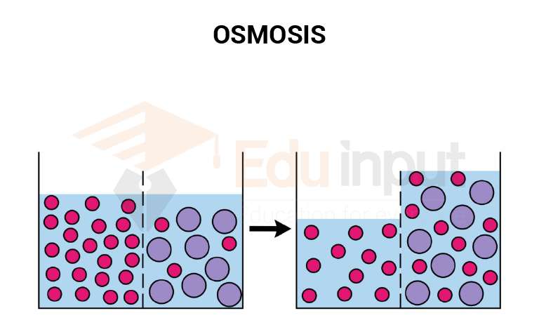
The loop of the nephron increases the efficiency of reabsorption by a countercurrent flow. Water is reabsorbed during countercurrent exchange. More water and ions are reabsorbed through the longer loop of the nephron. Thus desert rodents like kangaroo rats form highly concentrated urine. They have very long loops in the nephron. But amphibians live in aquatic habitats. So their nephrons lack a loop.
Countercurrent Exchange Mechanism
There are the following steps of the contour current mechanism.
1. Some salt (NaCl) and water are reabsorbed in the proximal convoluted tubule. It reduces its volume by approximately 25%. But salt and urea are still iso-osmotic with the extracellular fluid.
2. Now the filtrate moves to the descending limb of the loop of the nephron and loses water. Thus its volume is further reduced and it becomes more concentrated. There is a high salt concentration in the extracellular fluid. It is called a brine bath. So water moves out of the tubule by osmosis.
3. The highest urea brine bath concentration is present around the lower portion of the loop of the nephron. The filtrate passes into the ascending limb. Sodium (Na) ions are actively transported out of the filtrate into the extracellular fluid. Chloride ions follow it passively.
4. The cells of the ascending limb are impermeable to water. Therefore, water cannot flow out of the ascending limb. Thus, the salt concentration of the extracellular fluid becomes very high.
5. The salt flows passively into the descending loop. But they move out again in the ascending loop. It causes the recycling of salt through the loop and the extracellular fluid. It flows in the descending and ascending limbs in opposite directions. Thus a countercurrent gradient in salt is set up. A large amount of urea moves out of the collecting ducts. Therefore, the osmotic pressure of the extracellular brine bath increases.
6. Finally, the distal convoluted tubule opens into the collecting duct. The collecting duct is permeable for urea. The concentrated urea in the filtrate diffuses out into the surrounding extracellular fluid. The high urea concentration in the extracellular fluid with a high concentration of salt forms the urea-brine bath. It causes water to move out of the filtrate by osmosis.
7. Many peritubular capillaries surround each nephron. They collect water and return it to the systemic circulation.
8. The renal pelvis of the kidney form ureter. The ureter carries urine to a urinary bladder. The urinary bladder is a storage organ.
9. The Urine leaves the body through the urethra. It opens at the body surface at the end of the penis (in human males) or just in front of the vaginal entrance (In females). The urinary bladder fills with urine. Therefore, tension is produced in the muscle walls. This tension stimulates the response. This response relaxes sphincter muscles at the entrance of the urethra. This response is called urination.
Leave a Reply