The Spinal Cord-Structure, Working Mechanism, and Functions
 written by Sidra Batool
The spinal cord is a tubular structure made up of nervous tissue that carries signals between the brain and body. The spinal cord is a long bundle of nerves that extends from the base of the skull down through the vertebral column and into the sacral region. It carries signals between the brain and the rest of the body. It also contains motor neurons that control movement.
The spinal cord is responsible for transmitting information throughout the body.
Structure Of Spinal Cord
Its cross-section shows the following parts:
1. Neural Canal:
It is present in the center of the spinal cord. This canal contains cerebrospinal fluid.
2. Grey Matter:
The grey matter consists of cell bodies and dendrites. It controls reflexes in the spinal cord. The butterfly-shaped region of the brainstem contains the nerve cell bodies arranged in three grey columns.
3. Nerve Roots:
The ventral and dorsal roots of the spinal nerves are present on the spinal cord. These roots contain main motor and sensory fibers (axons and/or dendrites).
4. White Matter:
Its axons are covered by the whitish myelin sheath. Therefore, it is called white matter.
5. Meninges:
Three layers of protective membranes are called meninges. They surround the spinal cord. They are continuous with similar layers that cover the brain. These are:
(A) Dura Mater:
The outer layer is called dura mater. The dura mater is the tough, Protective outer layer, and the epidural space is the space between it and the surrounding bone of the vertebrae. This space is filled with adipose tissue and contains a network of blood vessels.
(b) Arachnoid
It is the middle layer. It is delicate. It connects to the innermost layer. The arachnoid mater is appropriately named for its open, spindly appearance. The space between these two layers is called the subarachnoid space and it’s filled with cerebrospinal fluid.
(C) Pia Mater
It is the innermost layer. The pia mater contains small blood vessels. It nourishes the spinal cord.
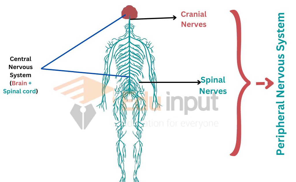
Spinal Nerves
The number of spinal nerves is directly related to the number of segments in the trunk and tail of a vertebrate. A frog has strong hind legs for swimming or jumping. It has a reduced trunk. It has no tail in the adult. It has only 10 pairs of spinal nerves. But a snake moves by lateral undulations of its long trunk and tall It has several hundred pairs of spinal nerves.
The spinal cord is a bundle of nerves and cells that extends from the brain to the lower back. The vertebral column is a series of bones that protect and support the spinal cord. The vertebrae are stacked on top of each other, from the pelvis to the skull. In between each pair of vertebrae, there is a spinal disk. These disks have a tough outer shell and a gel-like interior.
Working Mechanism
Signals from your brain to other body parts help you control your movements and involuntary functions like breathing and heartbeat. Your brain also gets information from other parts of your body about sensations like pressure or pain. The spinal cord also controls some reflexive (involuntary) movements without involving the brain. For example, the patellar reflex is managed by the spinal cord.
Functions of Spinal Cord
The spinal cord has two important functions in an animal. It is the connecting link between the brain and most of the body. It controls the spinal reflex actions. A reflex is a predictable, involuntary response to a stimulus. Reflexes control the movement of both voluntary and involuntary limbs.
One of its primary functions is transmitting signals from the brain to the body. This includes signals from the motor cortex, which is responsible for movement, and from the afferent fibers of the sensory neurons, which carry information about things like touch and pain.
Leave a Reply