Anatomy of Human Brain-Parts and Functions
 written by Sidra Batool
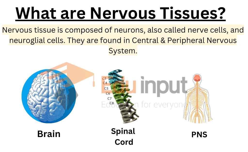
The central nervous system is like the control center for the body. It includes the brain, which is like the CEO, and the spinal cord, which is like the main line of communication. The peripheral nervous system is made up of all the other nerves that branch off from the spinal cord and brain.
These nerves are like different departments in a company, each with its specialty. The central nervous system includes the brain and spinal cord, while the peripheral nervous system is made up of nerves that branch off from the spinal cord and brain.
Also Read: What is Anatomy?
Brain of Vertebrates
The vertebrate brain develops at the anterior end of the spinal cord. The brain undergoes regional expansion during embryonic development. It formed from a hollow tube of nervous tissue. This tube develops into the hindbrain, midbrain, and forebrain. The central canal of the spinal cord extends up into the brain. Brian has chambers called ventricles. The ventricles are filled with cerebrospinal fluid.
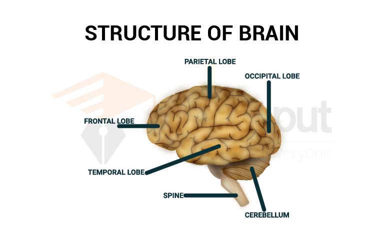
Parts of the Human Brain
There are three main parts of the human brain;
A. Hindbrain
The hindbrain is continuous with the spinal cord. It includes the medulla oblongata, cerebellum, and pons.
1. Medulla oblongata:
It is an enlargement of the spinal cord. It contains reflex centers for breathing, swallowing, cardiovascular function, and gastric secretion. It is well developed in all jawed vertebrates. It controls visceral functions. It screens for information that leaves or enters the brain.
2. Cerebellum:
It is an outgrowth of the medulla oblongata. It coordinates motor activity. This activity is associated with limb movement. It maintains posture and spatial orientation. The cerebellum is much larger in birds and mammals.
It shows a complex locomotory pattern in these animals. It also shows that these animals have a common evolutionary history of limb development and phylogeny.
3. Pons:
It is a bridge of transverse nerve tracts. It arises from the cerebrum of There forebrain to both sides of the cerebellum. It also contains tracts that connect the forebrain and spinal cord in all vertebrates.
B. Midbrain
Originally, the midbrain coordinates the visual input. It also took the functions of tactile (touch) and auditory (hearing) input. But its size has not changed. The roof of the midbrain is called the optic tectum. It is a thickened region of grey matter. It integrates visual and auditory signals.
C. Forebrain
The forebrain has two main parts: diencephalons and telencephalons or cerebrum;
1. Diencephalon
The diencephalon lies just in front of the midbrain. It contains the pineal gland, pituitary gland, hypothalamus, and thalamus.
Thalamus:
It relays all sensory information to higher brain centers.
Hypothalamus:
It lies below the thalamus. It regulates many functions like body temperature, sexual drive, carbohydrate metabolism, hunger, and thirst.
Pineal gland:
It controls somebody’s rhythms.
Pituitary gland:
It is a major endocrine gland. It secretes hormones.
2. Cerebrum
The cerebrum is present external to the corpus striatum. It has a large groove. This groove divides the cerebrum into the right and left cerebral hemispheres. The sensory and motor parts of the brain have changed with the evolution of vertebrates. Many functions shifted from the optic tectum to the cerebral hemispheres.
Thus it has increased the importance of the cerebrum. Therefore, it affected many other brain regions like the thalamus and cerebellum. The outermost part of the cerebrum is called the cerebral cortex. Different parts of the cerebrum have specific functions.
The cerebral cortex contains primary sensory areas and primary motor areas. Other areas of the cortex control the auditory signals from the environment. This includes the ability to use language in written and spoken forms humans.
Cranial Nerves
The peripheral nervous system of vertebrates is composed of paired cranial nerves and spinal nerves. Reptiles, birds, and mammals have 12 pairs of cranial nerves. Fishes and amphibians have the first 10 pairs.


Leave a Reply