Labeled Human Skeletal System Diagram
February 17, 2024  written by Sidra Batool
written by Sidra Batool Table of Contents
Human Skeletal System Diagram
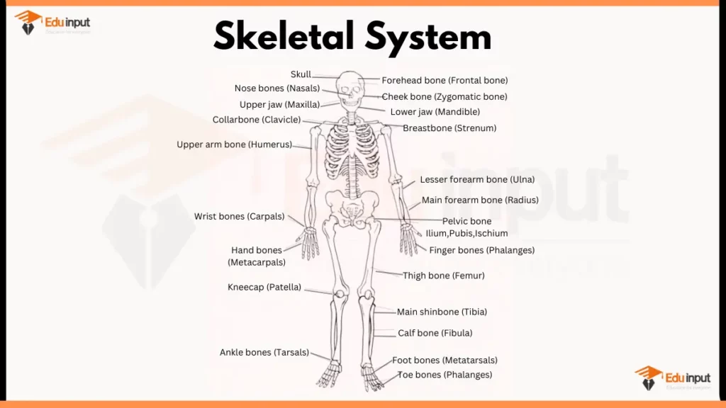
Here are the main parts of Human Skeletal System:
Axial Skeleton
- Skull: Protects the brain and houses sensory organs like eyes, ears, and nose.
- Cranium: Composed of eight bones fused together, forming the braincase.
- Facial bones: Fourteen bones that make up the face, including the jaw, cheekbones, and eye sockets.
- Vertebral column (spine): Provides support and flexibility. It protects the spinal cord.
- 24 vertebrae divided into five regions: cervical (neck), thoracic (chest), lumbar (lower back), sacrum (pelvis), and coccyx (tailbone).
- Intervertebral discs cushion the vertebrae and absorb shock.
- Rib cage: Encloses and protects the vital organs in the chest cavity (heart, lungs).
- 24 ribs attached to the thoracic vertebrae, some directly to the sternum (breastbone).
Appendicular Skeleton
- Pectoral girdle (shoulder): Connects the arms to the axial skeleton.
- Clavicle (collarbone) and scapula (shoulder blade) form the shoulder joint.
- Upper limbs
- Humerus (upper arm bone).
- Ulna and radius (forearm bones).
- Hand bones: carpals (wrist), metacarpals (palm), and phalanges (fingers).
- Pelvic girdle (hip): Connects the legs to the axial skeleton.
- Two innominate bones (each fused from ilium, ischium, and pubis) form the pelvis.
- Lower limbs
- Femur (thigh bone).
- Patella (kneecap).
- Tibia and fibula (lower leg bones).
- Foot bones: tarsals (ankle), metatarsals (sole), and phalanges (toes)
File Under:
Leave a Reply