Appendicular Skeleton | Blood Supply in Appendicular Skeleton
 written by Sidra Batool
The appendicular skeleton is the part of the skeleton of vertebrates that supports the appendages. The appendicular skeleton includes the skeletal elements within the limbs, as well as the supporting shoulder and pelvic girdles.
The upper and lower limbs form the appendicular skeleton. There are anatomical connections between the appendicular skeleton and axial skeleton, that are provided by the shoulder girdle and pelvis.
There are two bilateral joints where the appendicular skeleton is directly articulated with the axial skeleton.
Appendicular Skeleton
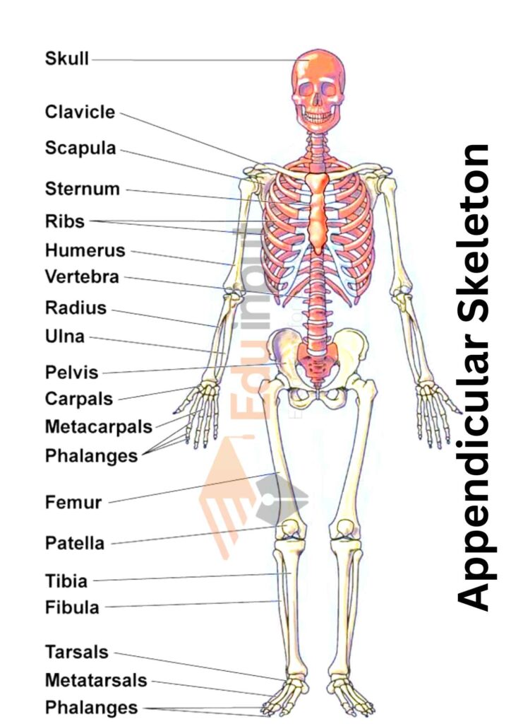
The appendicular skeleton consists of a pectoral girdle with appendages (fore limb) and a pelvic girdle with appendages (Hind Limb).
Pectoral Girdle
The pectoral girdle is composed of the scapula, Suprascapular, and clavicle. The clavicle connects the scapula with the sternum.
Fore Limb
The fore limb consists of one humerus, two radius and ulna, eight carpals, five metacarpals, and fourteen phalanges:
- Humerus: It is the proximal end that forms the ball and socket joint with the scapula. While its distal end form hinges joint with the radius and ulna.
- Radius and Ulna: They form multistage joints with eight wrist bones or carpals at their distal end.
- Carpals: There are 8 carpals. They form the wrist of the arm.
- Metacarpal: There are five metacarpal. They form the framework of the palm.
- Phalanges: There are 14 phalanges. They are attached to the metacarpals. They support the finger.
Pelvic Girdle
The pelvic girdle attaches the hind limb to the vertebral column. It consists of two Coxal bones. Each Coxal bone is formed by the fusion of three bones, Ilium, Ischium and Pubis.
The pelvic girdle supports the pelvic region.
Hind Limb
The hind limb consists of the following bones:
- Femur: There is a single femur in each leg. The proximal end of the femur forms the hip joint with the pelvic girdle or hip bone. It is a ball and socket joint. The distal end of the femur forms the knee joint with the proximal end of the tibia and fibula.
- Tibia and fibula: These are two in number in each leg. The distal end of the tibia and fibula form a joint with tarsal bones.
- Tarsals: There are eight tarsal which forms the ankle. The distal end of the tarsals is attached to the five metatarsals.
- Metatarsal: There are five metatarsals. They form the sole. Their distal ends are attached to the fourteen phalanges.
- Phalanges: There are five rows of fourteen phalanges of the toes. They are present in the fingers.
Blood Supply in Appendicular Skeleton
The lower extremity blood supply comes from the common iliac arteries, which are the terminal branches of the descending aorta.
The common Iliac artery divides into the internal and external iliac arteries, which supply all the structures of the pelvis and the lower extremities. The Subclavian artery supplies the arm and shoulder.
The right Subclavian artery is a branch of the Brachiocephalic trunk on the right or a branch directly off the aortic arch on the left.
Latest Research About Appendicular Skeleton
- Scientists have investigated whether muscle ultrasonography can estimate appendicular skeletal muscle mass (ASM) and screen for sarcopenia. Muscle thickness and echo-intensity in four muscles were measured in 212 volunteers, and ASM prediction equations were developed and validated. Muscle ultrasound may be effective for estimating ASM and screening sarcopenia. [1]
- Scientists have used a double staining method to investigate the early developmental characteristics of the skeletal system in Miichthys miiuy larvae. They systematically observed and described the morphological development of the spinal vertebrae and appendicular skeleton, and found spinal abnormalities and strong correlations between skeletal length and age, as well as total and body length. [2]
- Scientists developed and validated prediction equations for total body, arm, and leg skeletal muscle mass using whole-body MRI and DXA for 475 adults, providing practical tools for research and clinical settings. [3]
- Scientists describe the appendicular skeleton of Rinconsaurus caudamirus from Argentina, noting its unique features and phylogenetic relationships within titanosaurs. The estimated body mass is 3-5 tonnes, and it is classified as a member of the clade Rinconsauria. [4]

Leave a Reply