Structure of Nephron-Types, Blood Supply, and Function
 written by Sidra Batool
What is Nephron?
Nephrons are structural and functional units of the kidney. They are responsible for filtering and purifying blood. It plays important role in maintaining homeostasis by removing waste products, excess electrolytes, and fluids from the bloodstream to form urine.
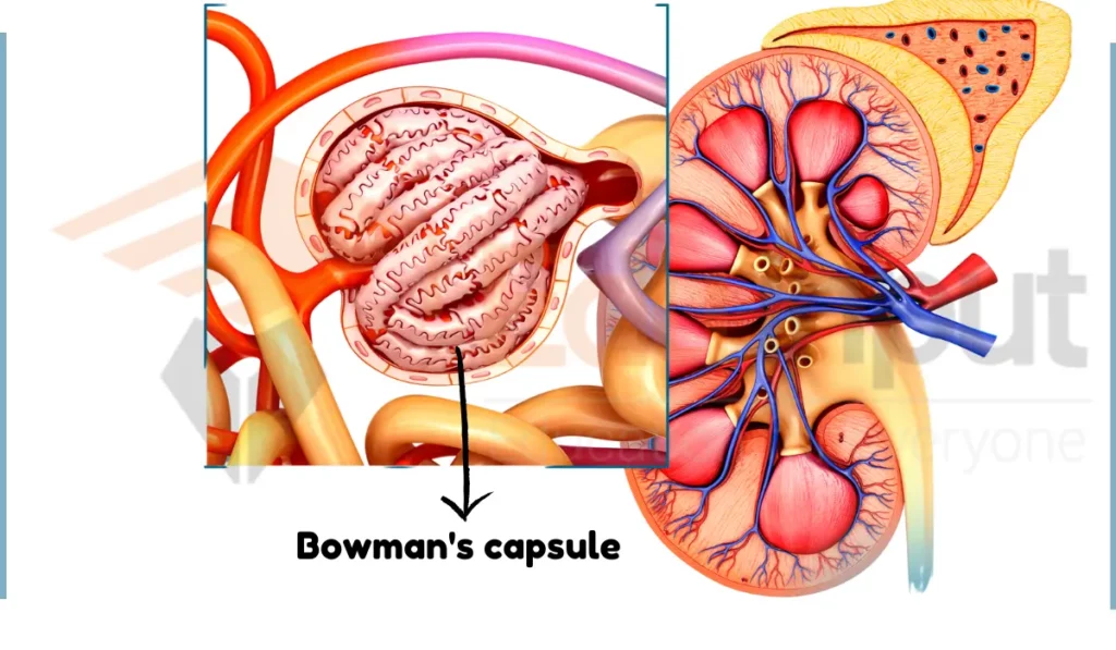
Glomerular endothelial cells, Glomerular basement membrane, and Podocytes form the filtration membrane in the nephron. Together, these three structures form a highly selective filtration barrier that allows the glomerulus to efficiently filter blood plasma into the Bowman’s capsule.
Types of Nephron
There are two main types of nephron based on their position:
1. Cortical Nephrons
Nephrons that are present along the cortex are known as cortical nephrons. They make 85% of all nephrons. These nephrons have their renal corpuscles. It is composed of glomerulus and Bowman’s capsule, as well as the initial segments of their tubules. These tubules are situated in outer cortex region of the kidney.
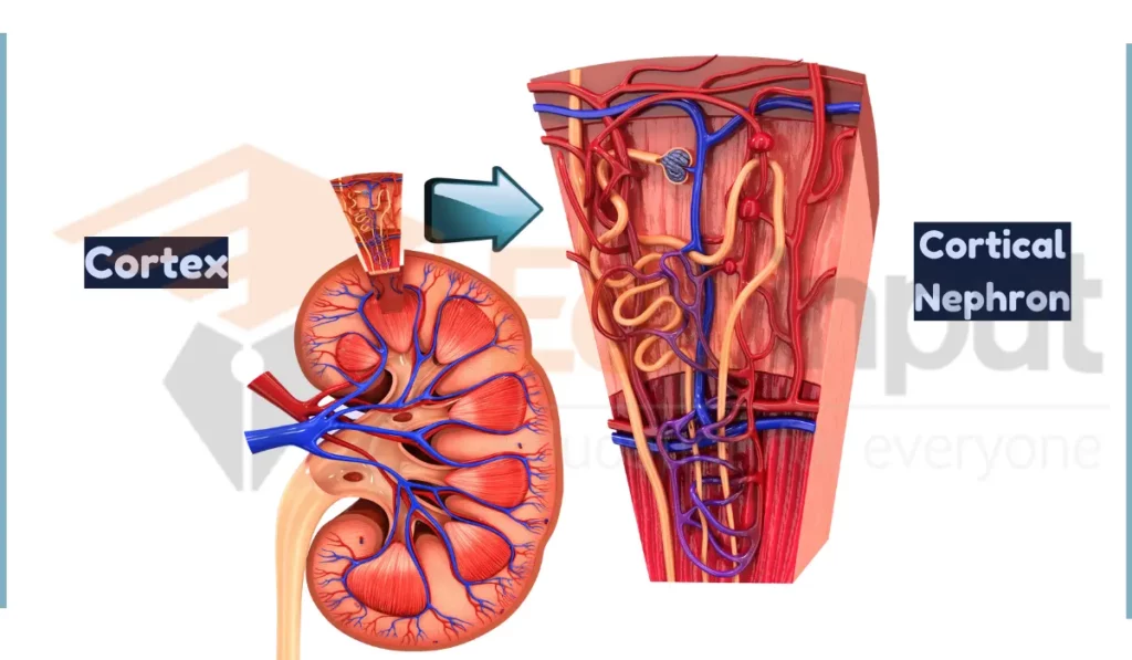
Cortical nephrons mainly perform excretory and regulatory. Cortical nephrons are involved in general filtration processes within the kidney. They are also involved in initial stages of urine formation by filtering blood. It also selectively reabsorbs essential substances.
2. Juxtamedullary Nephrons
Juxtamedullary nephrons are characterized by a tubular system that forms a loop extending deep into the inner medulla of the kidney.
They Make up about 15% of all nephrons. They specialize in concentrating and diluting urine. Their unique tubular loop facilitates efficient concentration of solutes in the renal medulla, contributing to the regulation of water balance and the formation of urine with varying levels of concentration.
Nephron Anatomy
Each nephron has the following structures:
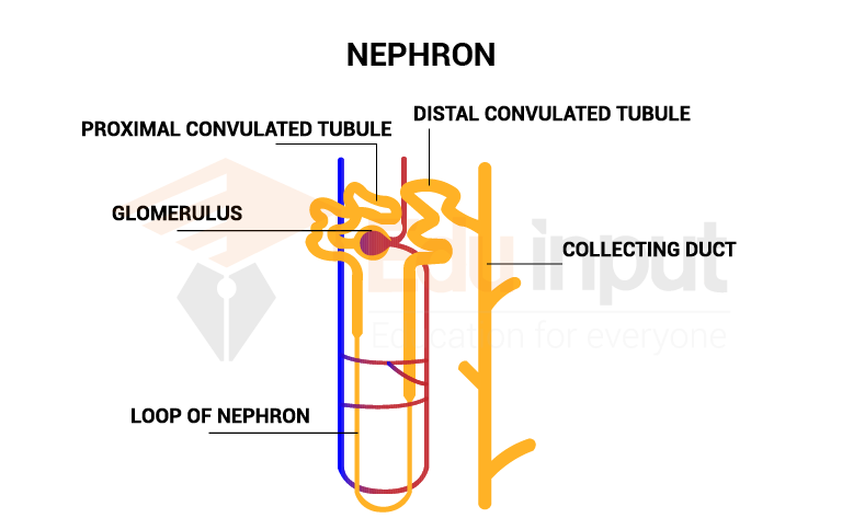
1. Bowman’s capsule
The long tube-like structure of the mammal’s nephron varies from 35 to 55mm long. At one end, the tube is closed, folded, and expanded into a cuplike structure called the Bowman’s capsule, which is also known as a renal capsule. Bowman’s capsule is a cup-shaped swelling present at the inner end of each nephron.
2. Proximal tubule
The proximal tubules are part of the nephrons, serving as the site where the glomeruli filter blood plasma and send it down the renal tubules. Most solute reabsorption in the nephron occurs in the proximal tubule.
The proximal tubules consist of two parts: the initial convoluted portion and the subsequent straight descending portion.
The initial convoluted portion contains the thick ascending limb of Henle’s loop. The subsequent portion is correctly termed the straight descending portion.
Functions
- Initial convoluted portion: This section reabsorbs essential nutrients and water from the filtrate back into the bloodstream, using various transport mechanisms. Glucose is primarily reabsorbed in the proximal convoluted portion of the proximal tubule in the nephron.
- Straight descending portion: This loop continues reabsorption of water and some electrolytes, helping concentrate the filtrate further.
3. Loop of Henle
The loop of Henle consists of two segments: the descending limb and the ascending limb. Both segments filter out specific substances. The salt level-monitoring part of the nephron is also associated with loop of Henle.
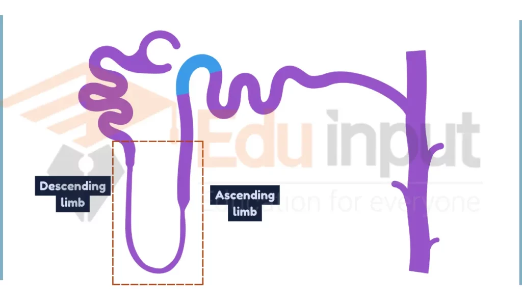
Functions
- Descending limb: Highly permeable to water, allowing it to passively move out into the surrounding tissue, concentrating the filtrate and creating a hypertonic medulla (inner region of the kidney).
- Ascending limb: Impermeable to water but actively pumps out sodium and chloride ions, preventing them from being reabsorbed, maintaining the filtrate’s dilution.
4. Distal tubule
The distal tubule, the final section of the nephron where urine passes through, connects and empties into the collecting ducts lining the medullary pyramid. These collecting ducts combine and join together to form the papilla, which collects urine from the nephron and transports it to the bladder.
Functions
- Fine-tune electrolyte and water balance in the filtrate under hormonal control (e.g., aldosterone and antidiuretic hormone).
- Further contributes to urine concentration by reabsorbing water in response to antidiuretic hormone.
5. Collecting tubules
The collecting tubules open into the pelvis of the kidney. Filtrate from the glomerulus takes place in these structures, and urine is formed.
Functions
- Collect the diluted or concentrated filtrate from the distal tubules of multiple nephrons.
- Final site for water reabsorption under antidiuretic hormone control, influencing urine volume and concentration.
- Empty the concentrated or diluted urine into the renal pelvis, which drains to the ureter and bladder.
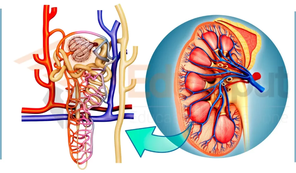
Blood Supply to Nephron
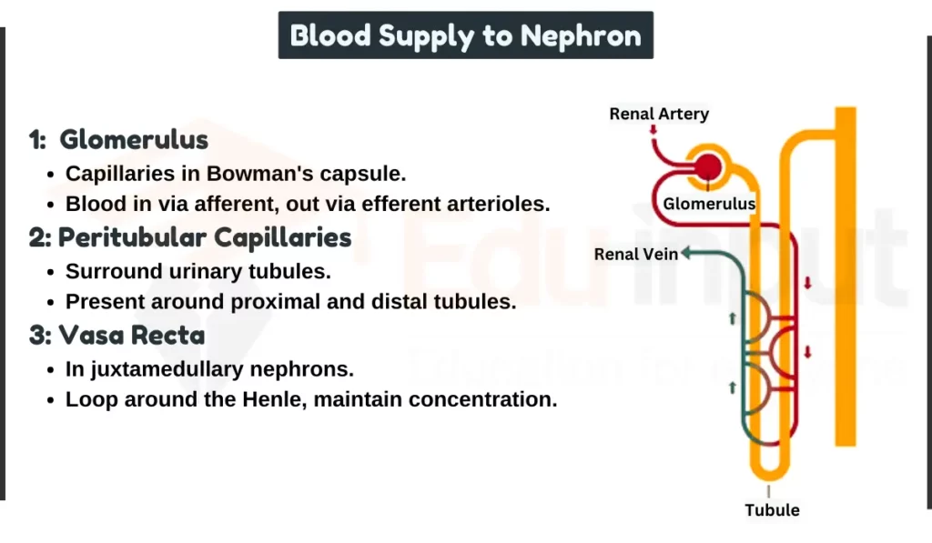
1. Glomerulus
Bowman’s capsule surrounds a ball of capillaries known as the glomerulus, facilitating blood circulation through the capsule in two steps:
- Glomerulus enters the capsule through afferent arterioles.
- It leaves the capsule through efferent arterioles.
2. Peritubular Capillaries
The blood vessel further subdivides into another network of capillaries known as peritubular capillaries. These capillaries surround all the urinary tubules, with a notable presence around the proximal and distal tubules.
3. Vasa Recta
In juxtamedullary nephrons, additional capillaries, called vasa recta, are present. These capillaries extend down to form a loop around the loop of Henle. The vasa recta play a crucial role in maintaining the concentration gradient in the renal medulla.
Regulation of Blood Flow
Autoregulation and hormonal control play pivotal roles in adjusting blood flow to different parts of the nephron. Autoregulation ensures a constant blood supply, while hormones like renin and angiotensin regulate blood pressure and volume.
Functions of Nephron
- Filtration: Primary function of nephrone is filtration. The glomerulus in the Bowman’s capsule filters blood plasma, creating a fluid called the filtrate. This filtrate contains essential molecules like water, glucose, and electrolytes alongside waste products.
- Selective Reabsorption: Different parts of the nephron tubules specialized cells reabsorb specific substances from the filtrate back into the bloodstream. Key examples include:
- Proximal tubule: Reabsorbs water, glucose, amino acids, and other essential molecules.
- Loop of Henle: Actively pumps out sodium and chloride ions, creating a concentration gradient in the medulla. This allows for further water reabsorption in the collecting ducts.
- Secretion: Some substances like excess potassium and hydrogen ions are actively secreted from the bloodstream into the filtrate in certain nephron segments, contributing to blood pH regulation.
- Urine Formation: As the filtrate flows through the tubules, water and essential molecules are reabsorbed, while waste products remain. The remaining fluid, now called urine, exits the nephron and eventually the body through the bladder.

Leave a Reply