Animal Cell Diagram Explained
February 13, 2024  written by Sidra Batool
written by Sidra BatoolLabelled Diagram of Animal Cell
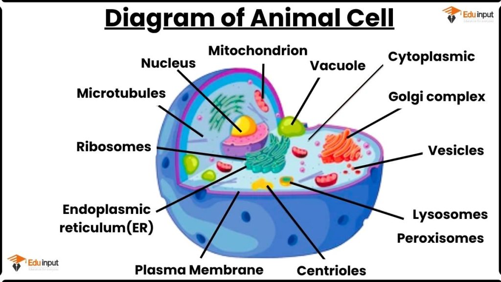
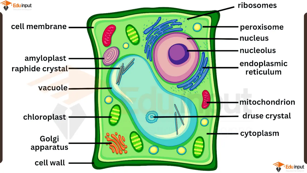
This Animal cell’s diagram shows following parts of the cell:
- Nucleus: This is the control center of the cell, and it contains the cell’s DNA. The nucleus is surrounded by a nuclear membrane.
- Cytoplasm: This is the jelly-like substance that fills the cell and contains all of the other organelles.
- Vacuole: This is a sac-like organelle that stores water, food, and waste products.
- Mitochondrion: These are the “powerhouses” of the cell, and they produce energy for the cell.
- Golgi complex: This organelle packages and distributes proteins and other molecules throughout the cell.
- Plasma membrane: This is the outer boundary of the cell, and it controls what enters and leaves the cell.
- Centrioles: These organelles help to organize the cell’s microtubules, which are important for cell division.
- Endoplasmic reticulum (ER): This organelle is involved in protein synthesis and transport.
- Ribosomes: These are tiny structures that make proteins.
- Lysosomes: These organelles break down waste products and old cell parts.
- Peroxisomes: These organelles break down harmful molecules.
File Under:
Leave a Reply