What Are Muscular Tissues?-Properties, Composition, Types, And Functions
 written by Sidra Batool
Muscular tissue is an animal tissue that works by contracting to apply forces to different parts of the body. It is made up of muscle cell fibers arranged in sheets and fibers, collectively called muscles. Muscles enable an organism to move and perform other contractile actions. Animals have three types of muscle tissue that differ slightly but work in a similar way.
Skeletal muscles make up a significant portion of an individual’s total body weight, typically accounting for 30 to 40% of the total mass. [1]
Muscle tissue is made up of cells called muscle fibers that can contract and produce movement. These cells are organized into bundles or layers and surrounded by connective tissue, with a rich supply of blood vessels. Muscle fibers contain contractile proteins called actin and myosin, which allow for muscle contraction. This unique composition and structure of muscle tissue makes it able to perform its functions in the body, including movement, support, and regulation of body functions.
What are Muscular Tissues?
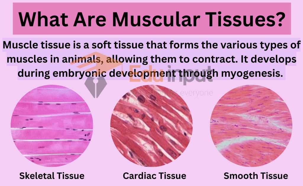
Muscle tissue is a soft tissue that forms the various types of muscles in animals, allowing them to contract. It develops during embryonic development through myogenesis. Muscle tissue is rich in contractile proteins, specifically actin and myosin, which facilitate movement by contracting and relaxing. In addition, muscle tissue contains regulatory proteins such as troponin and tropomyosin, among other muscle proteins.
Properties of Muscular tissues
Muscular tissue is composed of cells that possess unique properties that allow them to perform their functions. These properties are:
- Contractibility: ability of muscle cells to shorten forcefully, generating force for movement.
- Extensibility: ability of muscle cells to be stretched, accommodating movements and actions.
- Elasticity: ability of muscle cells to return to their original shape and length after being stretched.
- Excitability: ability of muscle cells to respond to stimuli from motor neurons or hormones, generating electrical signals that trigger muscle contraction.
Location of Muscular Tissues
The skeletal muscles are connected to bones and facilitate voluntary movements, while the smooth muscles are involved in involuntary functions such as regulating organ function and blood flow.
The cardiac muscles are found in the heart and responsible for pumping blood through contractions. In addition to their role in regulating organ function, smooth muscles are present in the eyes and help in focusing by changing the shape of the lens.
Smooth muscle is also responsible for peristalsis, which involves waves of contraction throughout the digestive system that aid in the movement of food through the body.
Composition & Structure of Muscular tissues
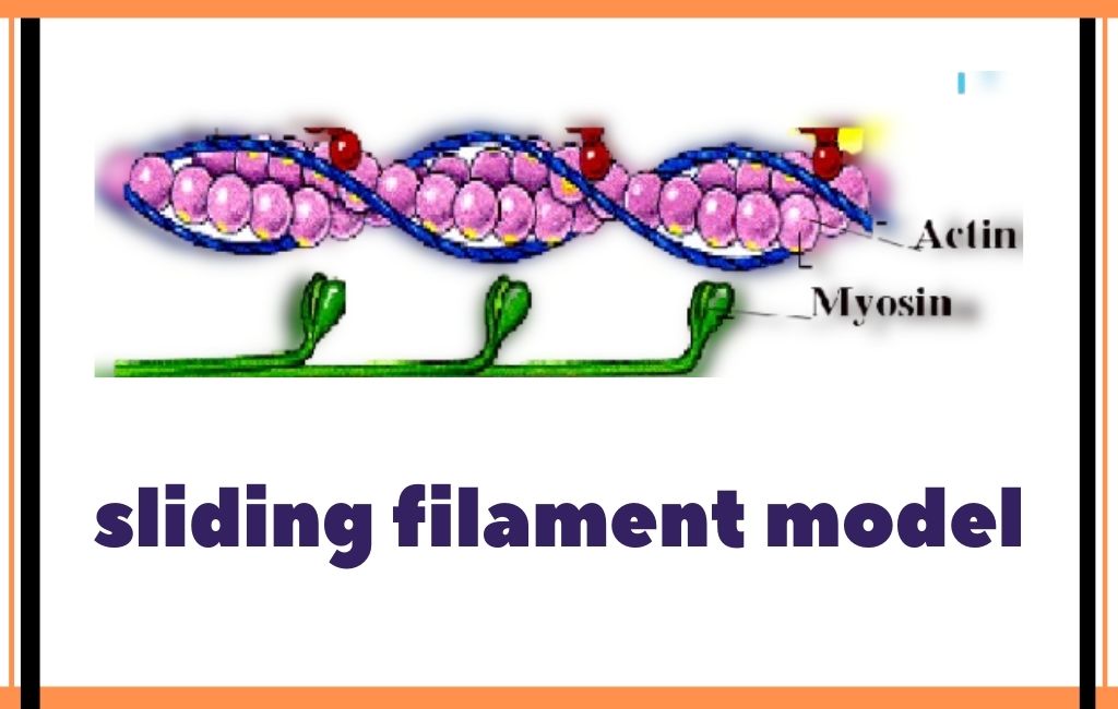
Muscular tissues is made up of elongated cells called muscle fibers. These are specialized for contraction. These muscle fibers contain contractile proteins such as actin and myosin. These muscle fibers are responsible for generating the force necessary for movement.
Muscle fibers are organized into bundles. These bundles are arranged in parallel form to make build muscle tissue. The arrangement of these bundles, fibers, and proteins, give rise to the different types of muscle tissue, such as skeletal, cardiac, and smooth muscle. Each type of muscle tissue has its own structure and function.
Along with contractile proteins, muscle tissue also contains regulatory proteins like troponin and tropomyosin, which helps in regulation of muscle contraction. These proteins work together to control the interactions between actin and myosin, allowing for precise control of muscle movement.
Muscle tissue also contains other proteins that contribute to muscle function and maintenance. For example, titin is a large protein that acts as a scaffold, providing structural support to muscle fibers. Myoglobin is a protein that stores oxygen in muscle tissue, helping to fuel muscle contraction.
Muscles are composed of thousands of fibers, which are surrounded by different types of coverings or sheaths. It contains:
- Epimysium: the outermost layer surrounding the entire muscle
- Perimysium: the middle layer surrounding bundles of muscle fibers
- Endomysium: the innermost layer surrounding individual muscle fibers
Types Of Muscular Tissue
There are three main types of muscular tissues, based on their composition and function.
- Skeletal muscle tissue
- Cardiac Muscle tissue
- Smooth muscle tissue
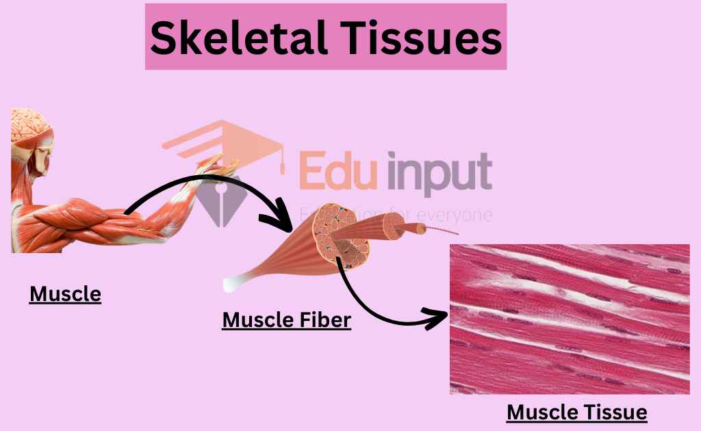
Skeletal Muscle Tissue
Skeletal muscle tissue is a specialized tissue characterized by its long, striated appearance. It can vary in length from several millimeters to 10 centimeters and typically measures 10 to 100 micrometers in width. The tissue is composed of bundles of myofibrils, containing contractile units known as sarcomeres, that give it the characteristic striped appearance.
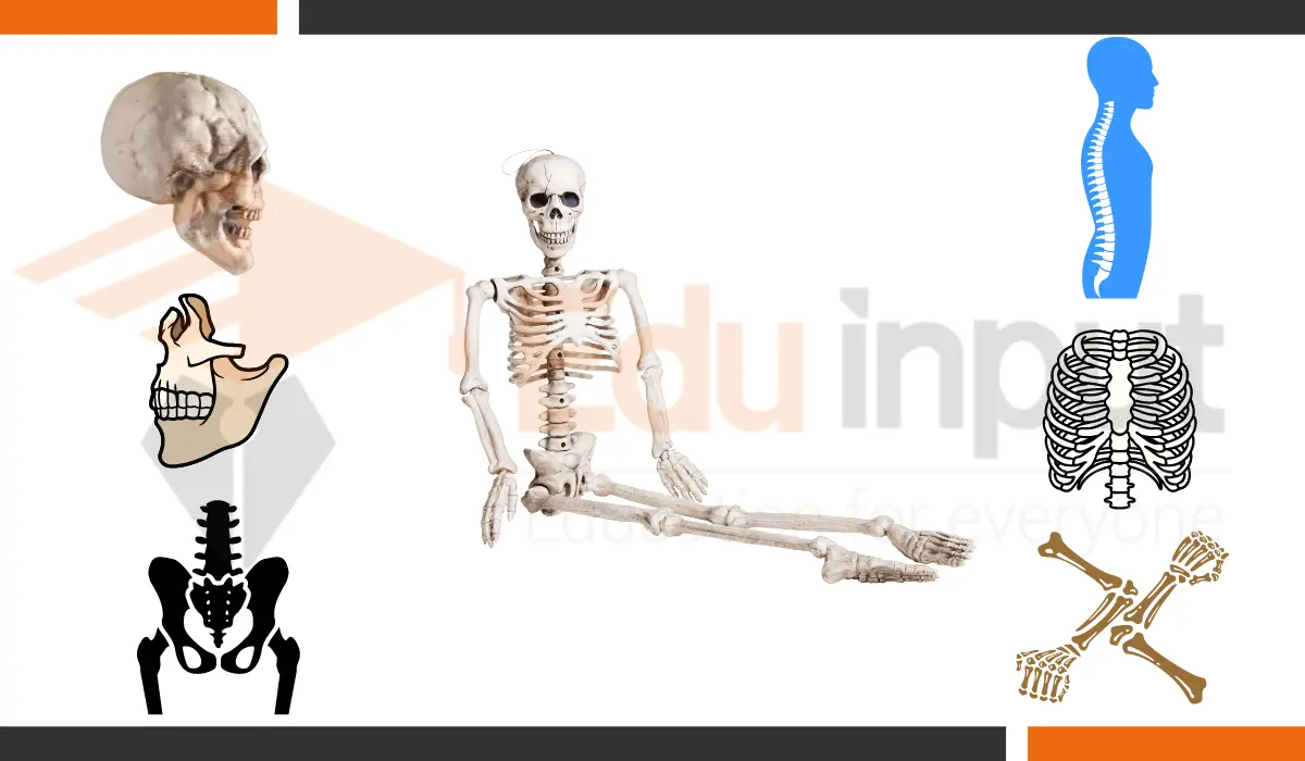
Skeletal muscle is attached to bones or aponeuroses by tendons. It is primarily responsible for voluntary movements, such as locomotion and maintaining posture.
Skeletal muscle is a type of striated muscle that makes up almost 40% of a human’s body weight. It is composed of long, cylindrical, and multi-nucleated cells that are formed by the fusion of embryonic myoblasts. Each nucleus in these cells regulates the metabolic requirements of the sarcoplasm around it. Due to their high energy requirements, skeletal muscle cells contain many mitochondria that generate ATP.
The striations in skeletal muscle tissue are formed by sarcomeres, highly organized bundles of actin, myosin, and associated proteins. These organized bundles allow for quick muscle contractions and release. Skeletal muscle tissue is attached to bones through tendons, which are highly elastic portions of connective tissue. While many muscles may appear to control a single appendage, in reality, each muscle only controls one small aspect of movement. Skeletal muscle tissue can be voluntarily controlled by the somatic nervous system.
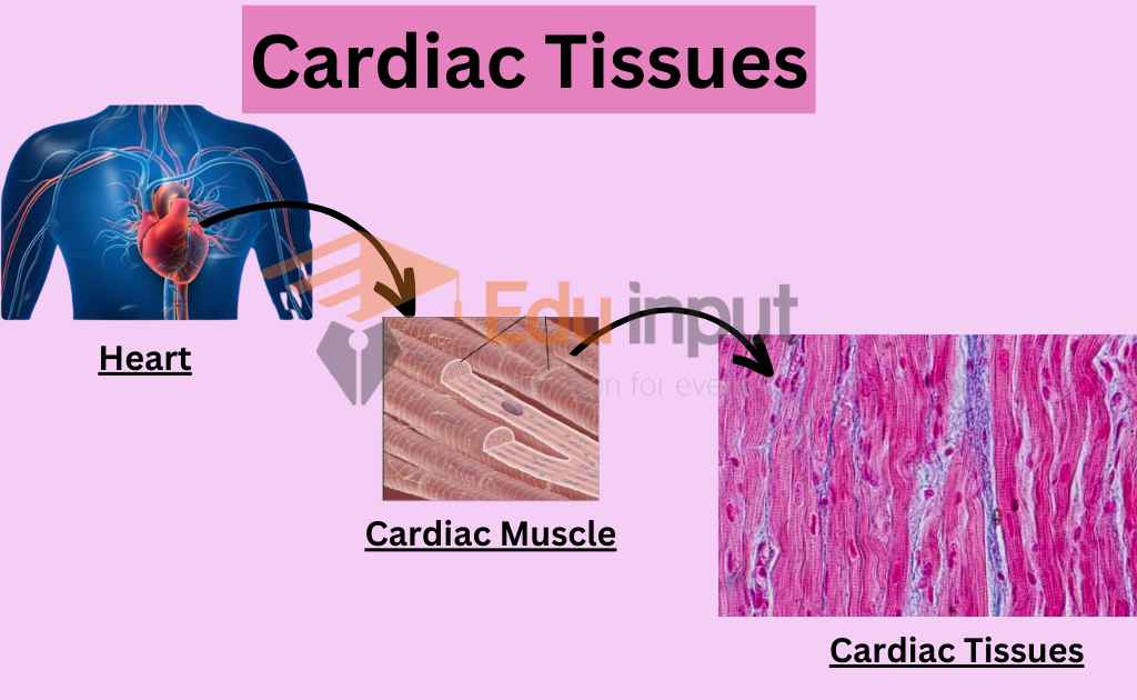
Cardiac Muscle Tissue
Cardiac muscle is a specialized type of muscle tissue found only in the walls of the heart as myocardium. Cardiac muscle tissue has complex and branching striations. These branching cells are connected via intercalated discs. These discs help cardiac muscle cells to contract as one and provide a rapid and coordinated contraction to move blood.
Cardiomyocytes, the cells that make up cardiac muscle tissue, are short and narrow, and fairly rectangular in shape. They contain many sarcosomes, which provide the required energy for contraction. Unlike skeletal muscle cells, they normally contain a single nucleus. Cardiomyocytes generally contain the same cell organelles as skeletal muscle cells, although they contain more sarcosomes.
Cardiomyocytes are large and muscular and are structurally connected by intercalated discs, which have gap junctions for diffusion and communication. These unique aspects of cardiomyocytes allow the transmission of contractile force between cells as electrical depolarization propagates from cell to cell.
Cardiomyocytes generate enough contractile force for the heart to beat effectively. They contract together in unison, causing enough pressure to force blood around the body.
The heart’s atria and ventricles propel blood to the left/body/systemic and right/lungs/pulmonary circulatory systems through coordinated contractions of cardiac muscle cells. This process exemplifies systole in the heart.
Cardiac muscle cells require a steady supply of oxygen and nutrients, as well as the removal of waste products such as carbon dioxide, through an adequate blood and electrical supply.
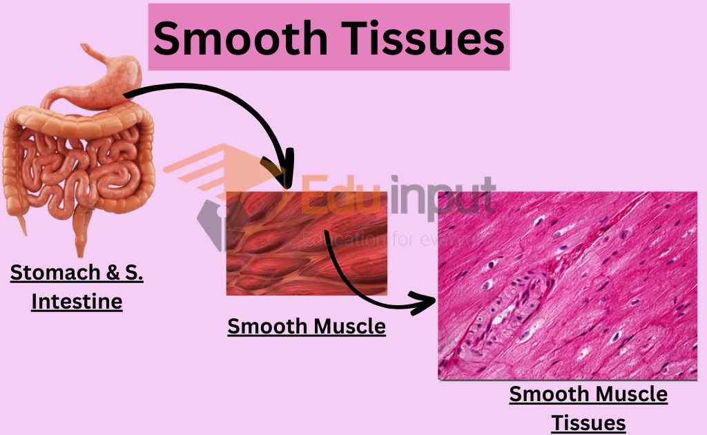
Smooth Muscle Tissue
Smooth muscle fibers are located in the walls of hollow visceral organs and blood vessels, and are under involuntary control. They appear spindle-shaped, lack striations, and have thin, randomly arranged actin and myosin filaments. Smooth muscle is divided into single-unit and multiunit subgroups, and innervates either whole bundles or individual cells.
It is found in various organs, including the urinary bladder, gastrointestinal and respiratory tracts, and the iris of the eye. While the structure and function of smooth muscle cells are similar across organs, the stimuli that induce contraction are different.
Single-unit smooth muscle cells are arranged in bundles or sheets, and when they contract, they do so as a syncytium, meaning that the entire bundle or sheet contracts together. This type of smooth muscle is found in the walls of many visceral organs such as the stomach, intestines, and uterus.
Multiunit smooth muscle, on the other hand, consists of individual cells that are innervated and controlled separately. This type of smooth muscle is found in the iris of the eye and in the walls of blood vessels.
Functions of Muscular Tissues
Muscular tissue is responsible for generating force and enabling movement in animals. The main functions of muscular tissue include:
- Movement: Muscles enable an organism to move and perform other contractile actions.
- Support: Muscles provide structural support to the body and help maintain posture.
- Regulation: Smooth muscles are involved in involuntary functions such as regulating organ function and blood flow. Cardiac muscles are responsible for pumping blood through contractions.
- Heat production: Muscles generate heat during contraction, which helps maintain body temperature.
- Protection: Muscles protect internal organs and tissues by cushioning them from external forces.
- Peristalsis: Smooth muscle aids in the movement of food through the digestive system through waves of contraction known as peristalsis.
Modern Research In field of Muscular Tissues
- Stem cell protocols allow for the engineering of human skeletal muscle tissue in vitro using various techniques like cell cultures, microfluidics, organoids, and bioprinted constructs. These models offer different levels of complexity and are useful for drug discovery, with several drugs already tested. More advanced skeletal muscle tissue models are being developed for personalized medicine and disease modeling. [2]
- A study investigated surface instability of active-elastic skeletal muscle tissue subject to shear. Two models were considered, and an activation parameter was introduced to describe microstructural contraction. Results showed a decrease in critical shear with an increasing ratio, and a decrease in stability with an increase in anisotropic variant. A decay deformation tensor was also introduced to model muscle mass loss during sarcopenia. The second model was found to be more compatible with decreasing stability with muscle mass loss. [3]
- Studies investigated the effect of alfalfa protein concentrate (APC) on turkey muscles. The use of APC in the diet stimulated antioxidant processes, improving the antioxidant status of turkeys. Continuous use of APC proved to be a better feeding practice to optimize oxidative potential than periodic inclusion. [4]

Leave a Reply