How the cross-bridges are controlled? | Role of Ca2+in Skeletal Muscle Thin Filament | Energy for muscle contraction
 written by Sidra Batool
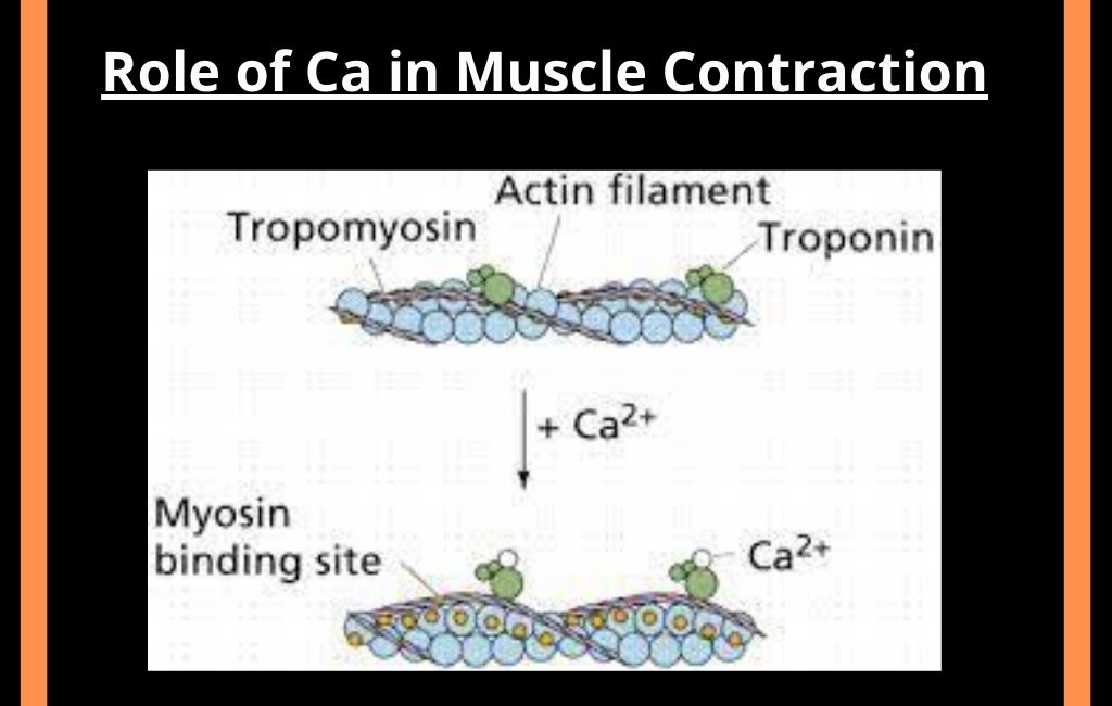
The role of Ca2+ in the contraction of skeletal muscle is very critical in the activation and deactivation of contractile proteins. Once the binding sites are exposed, the myosin binds to actin to begin cross-bridge cycling by using ATP. Then the sarcomere shortens and the muscle contracts. The presence of free calcium is an essential factor in muscle contraction. As binding does not occur in the absence of Ca.
At rest
The tropomyosin covers the sites on the actin chain during rest. Thus the head of the myosin cannot be attached to these sites.
During contraction
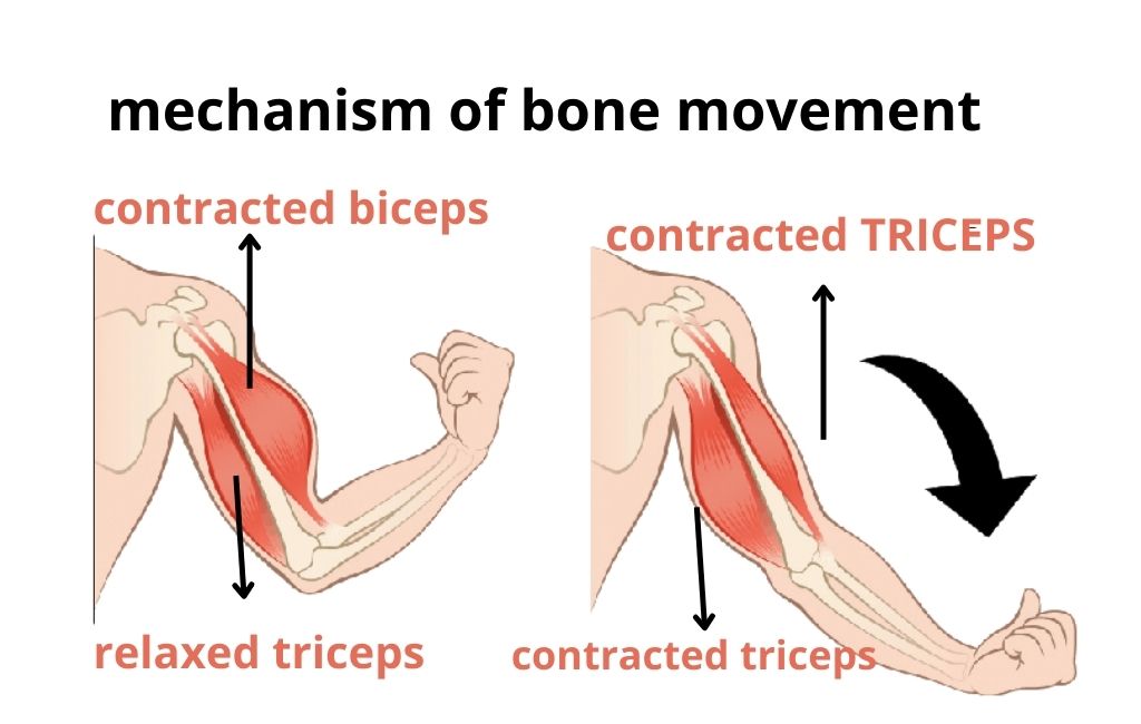
Calcium ions bind with the troponin molecule at the start of muscle contraction. The troponin molecule moves slightly. This movement displaces tropomyosin. It exposes the binding sites for the myosin head. The myosin head is attached to the actin filament. Atp is hydrolyzed and sliding of actin and myosin starts.
This ATP is provided by a large number of mitochondria present in each muscle cell.
How the actin-myosin interaction is controlled by ca++ ions?
The following process takes place during the acting in myosin interactions:
- The impulse arrives at the neuromuscular junction. It initiates muscle contraction. A single motor neuron innervates all the muscle fibers. It forms a motor unit.
- The sarcolemma of muscle fiber cells penetrate deep into the cell. It forms a hollow elongated tube, called the transverse tubule or t tubule. The lumen of the t tubule is continuous with the extracellular fluid. Thousands of t tubules of each muscle cell are collectively called the t system.
- T tubule extends and encircles the mesophilic at the level of the z line or a and I junction.
- T tubule and the terminal portion of the adjacent envelope of sarcoplasmic reticulum from the triads. Triads are present at regular intervals along the length of the fibril.
- The nerve impulse is carried through the t tubule to the adjacent sarcoplasmic reticulum. The calcium gates of the SR open and release calcium into the cytosol .these calcium ions bind to the troponin molecule of the thin filament.
The binding site is exposed. Thus the cross disease of myosin attaches with the actin and contraction occurs.
All or none response
The contraction of each muscle is based on all or non-principle. All of its fibers participate in contraction. The degree of contraction depends upon the number of fibers that participate in contraction
Energy for muscle contraction
There are the following sources of energy for muscle contraction
ATP
Energy for muscle contraction comes from ATP. ATP is produced by the aerobic breakdown of glucose in muscle cells glucose comes from stored glycogen in the cell.
Creatine Phosphate
Sometimes more energy is required due to high metabolism. it is provided by another energy-storing substance called creating in phosphate
Lectic Acid Formation
Sometimes lessen number of ATPs are produced due to oxygen deficiency or very high metabolism. ATP requirements are made by the anaerobic breakdown of glucose intellective acid lactic acid accumulation their energy is used to change the remaining lactic acid into glucose.
Related FAQs
How cross-bridges are controlled during muscle contraction?
Muscle contraction is regulated by the actin and myosin filaments sliding past each other. Cross-bridges that come from myosin filaments and attach to actin filaments during ATP hydrolysis are thought to manage this process.
What prevents cross-bridge formation?
Tropomyosin is a protein that helps to prevent contraction in a muscle without nervous input by blocking myosin binding sites on actin molecules. This prevents cross-bridge formation, which is necessary for muscle contraction.
Which molecules regulate cross-bridge attachment activity?
Proteins that regulate cross-bridge formation, such as troponin and tropomyosin, play an important role in muscle contraction.
Leave a Reply