Human Circulatory System Diagram Explained
February 17, 2024  written by Sidra Batool
written by Sidra Batool Table of Contents
Human Circulatory System Diagram
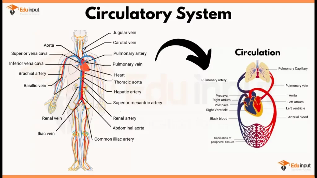
Here are key components of human circulatory system and their functions:
1. Heart
The heart has four chambers:
- Right atrium: Receives deoxygenated blood from the body.
- Right ventricle: Pumps deoxygenated blood to the lungs.
- Left atrium: Receives oxygenated blood from the lungs.
- Left ventricle: Pumps oxygenated blood to the body.
2. Blood Vessels
- Arteries: Carry oxygenated blood away from the heart. They have thick, muscular walls to withstand high pressure.
- Veins: Carry deoxygenated blood back to the heart. They have thinner walls and valves to prevent backflow.
- Capillaries: Tiny, thin-walled vessels where gas and nutrient exchange occurs between blood and tissues.
3. Blood
- Plasma: Liquid portion carrying dissolved nutrients, hormones, and waste products.
- Red blood cells (RBCs): Transport oxygen from lungs to tissues and carbon dioxide back.
- White blood cells (WBCs): Play a crucial role in the immune system, fighting infections.
- Platelets: Aid in blood clotting to prevent excessive bleeding.
File Under:
Leave a Reply