Human Digestive System Diagram Explained
February 14, 2024  written by Sidra Batool
written by Sidra BatoolHuman Digestive System Diagram
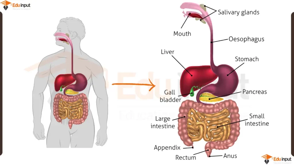
The digestive system is a complex series of organs that work together to break down food into nutrients that the body can absorb. The main parts of the digestive system are:
- Mouth: The mouth is where food enters the digestive system. Teeth break down food into smaller pieces, and saliva mixes with food to start the process of digestion.
- Esophagus: The esophagus is a muscular tube that carries food from the mouth to the stomach.
- Stomach: The stomach is a muscular sac that stores and churns food. Gastric juices in the stomach break down food into a liquid mixture called chyme.
- Small intestine: The small intestine is a long, narrow tube where most of the digestion and absorption of nutrients takes place. The small intestine is lined with millions of tiny villi, which increase its surface area and allow for better absorption of nutrients.
- Liver: The liver is a large organ that produces bile, a substance that helps to break down fats in the small intestine. The liver also stores nutrients, detoxifies the blood, and produces many other important chemicals.
- Gallbladder: The gallbladder stores bile produced by the liver.
- Pancreas: The pancreas produces enzymes that help to break down carbohydrates, proteins, and fats in the small intestine. The pancreas also produces insulin and glucagon, hormones that help to regulate blood sugar levels.
- Large intestine: The large intestine is a long, wide tube that absorbs water from the remaining food material and forms stool. Stool is then eliminated from the body through the rectum and anus.
File Under:
Leave a Reply