Dicots-Definition, Examples, Leaf and Root Morphology
 written by Sidra Batool
Dicotyledons or Dicots are a group of plants that have two embryonic leaves or cotyledons in their seeds. They are one of the two main groups of flowering plants, with around 200,000 different species.
The other group is called monocotyledons or monocots, and they usually have only one cotyledon. In the past, these two groups were considered as separate divisions of flowering plants.
Learn Difference Between Monocots and Dicots
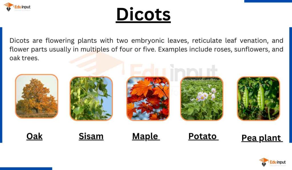
Examples of Dicots
Here are 20 examples of dicots:
- Peas
- Beans
- Daisy
- Lentils
- Peanuts
- Tomatoes
- Mint
- Oak
- Lettuce
- Apples
- Mangoes
- Oranges
- Cashews
- Magnolias
- Roses
- Geraniums
- Soybeans
- Carrots
- Sunflowers
- Buttercups
Leaf Venation in Dicots
Dicotyledonous plants have a distinctive leaf venation pattern that sets them apart from monocots. This venation pattern refers to the arrangement of veins within the leaf.
In dicot leaves, the vascular structures form a net-like pattern of veins, known as reticulate venation. This pattern arises from the branching and interconnecting veins that radiate from the central midrib.
The reticulate venation creates an intricate network, facilitating the distribution of water, minerals, and nutrients throughout the leaf.
This vein arrangement is in contrast to the parallel venation commonly found in monocot leaves.
Leaf Shape in Dicot Plants
Dicotyledonous plants exhibit a diverse range of leaf shapes, offering variations in surface area and photosynthetic capabilities. Unlike monocots, which often have linear or strap-like leaves, dicot leaves can take on numerous forms and sizes.
Dicot leaves commonly display ovate, lanceolate, or palmate shapes. Ovate leaves have a broad, rounded base that tapers to a point at the tip, resembling the shape of an egg.
Lanceolate leaves are elongated and narrow, gradually tapering to a point. Palmate leaves are characterized by multiple lobes radiating from a central point, resembling the shape of a hand with fingers spread out.
Leaf Shape and Photosynthetic Efficiency
The varied leaf shapes in dicotyledonous plants offer advantages in terms of surface area and exposure to sunlight. The larger surface area allows for more efficient light capture and photosynthesis, contributing to the plant’s overall energy production.
The diverse leaf shapes also aid in reducing water loss through transpiration and optimizing the distribution of resources within the leaf.
Leaf Morphology in Dicots
The morphology of dicot leaves reveals a dorsiventral structure, consisting of an upper surface called the adaxial surface and a lower surface called the abaxial surface. These two surfaces exhibit distinct differences in appearance and structure.
Epidermis and Cuticles
The epidermis is present on both the upper and lower surfaces of a dicot leaf. It consists of thin cuticles that provide protection against mechanical and physical damage.
Mesophyll
Between the upper and lower epidermis lies a layer called the mesophyll. It is composed of parenchymatous cells and contains chloroplasts responsible for photosynthesis.
The mesophyll can be classified into two types: palisade parenchyma and spongy parenchyma. The palisade parenchyma is located towards the upper surface (adaxial) and consists of elongated cells arranged parallel to each other.
On the other hand, the spongy parenchyma extends towards the lower epidermis and is composed of loosely arranged, round-shaped cells. These cells are interspersed with air cavities.
Stomata Distribution
The abaxial surface of dicot leaves generally contains a higher density of stomata compared to the adaxial surface.
Stomata are tiny openings that allow for gas exchange, including the intake of carbon dioxide and the release of oxygen and water vapor.
Vascular Bundles and Bundle Sheath Cells
The vascular tissues are organized into vascular bundles primarily located within the veins and midrib region of dicot leaves. These vascular bundles are protected by bundle sheath cells, which consist of one or more layers of parenchyma cells.
Root Morphology of Dicots
Dicot roots exhibit a taproot structure. It is characterized by a single, thick primary root that extends deep into the soil. The structure and arrangement of tissues in a dicot root can be observed at the cross-section of the root.
Rhizodermis or Epiblema
The outermost layer consists of a single layer of parenchymatous cells without intercellular spaces. It lacks stomata and cuticles. Root hairs are typically single-celled.
Cortex
The cortex is composed of loosely arranged parenchymatous cells that are oval or rounded in shape. These cells may serve as storage sites for food reserves.
Endodermis
The endodermis is made up of a single layer of barrel-shaped parenchymatous cells. The radial and inner tangential walls of endodermal cells are thickened with a substance called suberin, forming Casparian strips. However, these Casparian strips are absent in the endodermal cells that are located opposite to the protoxylem elements.
Stele
All the tissues present inside the endodermis make up the stele, which includes the vascular tissues.
Pericycle
The pericycle is generally a single layer of parenchymatous cells located internal to the endodermis. Lateral roots originate from the pericycle.
Vascular System
The vascular tissues are arranged radially. The tissue that separates the xylem and phloem is known as conjunctive tissue.
The xylem exhibits exarch and tetrarch conditions, and the metaxylem vessels are predominantly polygonal in shape.
Vascular System of Dicots
Dicot roots exhibit distinct characteristics. The xylem bundles are surrounded by phloem clusters, separated by the vascular cambium, which produces secondary growth.
The epiblema, or rhizodermis, lacks stomata and cuticles but produces single-cell root hairs. The cortex consists of thin-walled parenchymatous cells responsible for transporting salts and water.
The endodermis has barrel-shaped cells with passage cells and Casparian strips for regulating water and mineral movement.
The pericycle, a layer of parenchymatous cells, allows for lateral root development. Vascular bundles are arranged radially, with xylem containing tracheids, vessels, fibers, and parenchyma.
The phloem consists of sieve tubes, parenchyma, and companion cells. The central pith stores food reserves.
Latest Research About Dicots
- Scientists have made significant progress in understanding the molecular genetics and regulatory mechanisms of storage protein synthesis in seeds, exploring evolutionary origins, developmental processes, regulatory networks, and genetic modifications to enhance storage protein accumulation for improved nutritional quality. [1]
- Scientists have made significant progress in addressing severe global public health issues caused by micronutrient deficiencies through the development of biofortified rice varieties, particularly focusing on dicot ferritins and the rice nicotianamine synthase 2 (OsNAS2) gene, which have the potential to alleviate inadequate nutrient intake by up to 30-50%. [2]
- Scientists have developed a scientific literacy-based video quiz (VIDKU) utilizing the Sparkol Videoscribe application, incorporating learning videos and quizzes to enhance understanding of dicot and monocot materials, promoting scientific literacy through audio-visual media. [3]
Leave a Reply