What Are Tissues?-History, Types, And Examples
 written by Sidra Batool
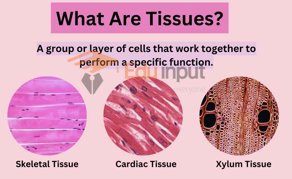
Tissues are groups of cells that work together to perform specific functions in our body. The intercellular matrix fills the spaces between cells. It’s like the “glue” that holds cells together within a tissue. Depending on the type of tissue, the matrix can vary in its composition and quantity.
Key Points
- Tissues are groups of cells that work together to perform specific functions in our body.
- The intercellular matrix is a non-living substance that fills the spaces between cells and holds cells together within a tissue.
- Tissues are hierarchical levels of organization between individual cells and complete organs, and assemblies of similar cells and their extracellular matrix from the same origin that together carry out a specific function.
- Tissues are absent from unicellular organisms and play an important role in regulating an organism’s response to its environment.
- The development and differentiation of tissues is a complex and highly regulated process that involves the expression of specific genes and the interaction of cells with each other and with their environment.
- The study of tissues and their structures has a rich history that dates back to the 19th century, and histology emerged as a distinct field of study concerned with the examination of tissues and their structures in the early 20th century.
- In animals, tissues are groups of similar cells and their intercellular material that work together to carry out a specific function. There are four main types of animal tissues: connective, muscle, nervous, and epithelial.
- Plants have various tissue types with unique functions: vascular, ground, epidermal, meristematic, and permanent tissue, each specialized to perform essential roles such as transport, support, protection, growth, and specialization.
What Are Tissues In Biology?
In Biology, tissues are considered to be a hierarchical level of organization between individual cells and complete organs. They are historically derived from the observation that groups of similar cells working together often have a common function. This has helped to define a tissue as assemblies of similar cells and their extracellular matrix from the same origin that together carry out a specific function.
Tissues are an important level of organization in multicellular organisms. Tissues are absent in unicellular organisms because they are composed of only one cell. Even among the simplest multicellular organisms, such as sponges, tissues are either poorly differentiated or completely lacking.
Tissues play an important role in regulating an organism’s response to its environment. For example, in animals, the nervous tissue is responsible for processing and transmitting information, while muscle tissue is responsible for movement. In plants, meristematic tissue is responsible for growth, and vascular tissue helps transport water and nutrients throughout the plant.
The development and differentiation of tissues is a complex and highly regulated process that involves the expression of specific genes and the interaction of cells with each other and with their environment.
History of Tissues
The study of tissues and their structures has a rich history that dates back to the 19th century. In 1801, Xavier Bichat introduced the term “tissue” to the field of anatomy, proposing that it is a fundamental component of human anatomy. He viewed organs as collections of different tissues, rather than as distinct entities.
Further developments in the study of tissues came with the understanding of embryology. It was discovered that all cells and tissues in the body derive from three germ layers in the embryo: the ectoderm, mesoderm, and endoderm. This realization laid the foundation for comprehending the development and differentiation of various types of tissues.
Gottlieb Haberlandt was a German botanist, who is recognized as the pioneer of plant tissue culture and is commonly referred to as the father of tissue culture. He was the first to isolate and cultivate plant cells using Knop’s salt solution. He is known for his groundbreaking work in separating and culturing plant cells using Knop’s salt solution.
Haberlandt was the first to demonstrate that plant cells are totipotent, meaning they can differentiate into any type of plant tissue. His work laid the foundation for the establishment of tissue culture as a vital tool in both basic and applied plant science research. [1]
In the early 20th century, histology emerged as a distinct field of study, concerned with the examination of tissues and their structures. The link between histology, anatomy, and physiology became apparent as it was found that the structure of each tissue in the human body is intricately connected to its specific functions.
What is Histology?
Histology is a branch of Biology that focuses on the study of the microscopic structure of tissues and organs in plants, animals, and humans. It involves the use of specialized laboratory techniques and instruments to examine tissues and cells at high magnification to identify their cellular components, organization, and functions.
Tissues In Animals
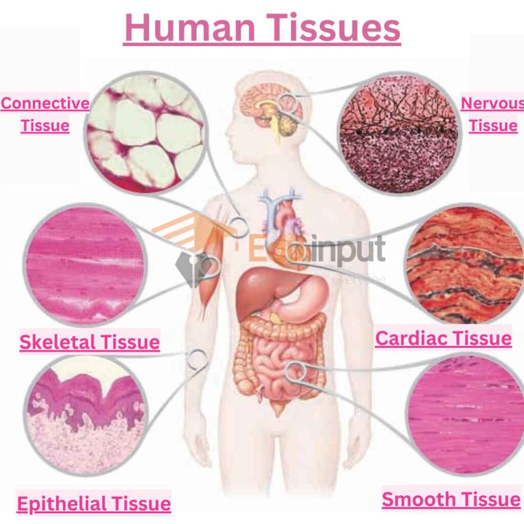
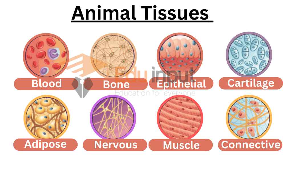
In animals, tissues are groups of similar cells and their intercellular material that work together to carry out a specific function. There are four main types of animal tissues: connective, muscle, nervous, and epithelial.
Connective Tissue
Connective tissue is found throughout the body, connecting and separating groups of other tissues and organs. It is made up of cells and ground substance, a gel-like material that surrounds cells, and most connective tissue contains fibers.
Fibers can be collagenous, elastic, or reticular, and allow for the diffusion of oxygen from blood vessels into cells. Connective tissue disorders include sarcomas, Marfan syndrome, lupus, and scurvy, which is a Vitamin C deficiency leading to fragile connective tissue.
Examples of Connective tissues: Tissues in tendons, ligaments, Blood, cartilage, bone, and adipose tissue.
Muscular Tissue
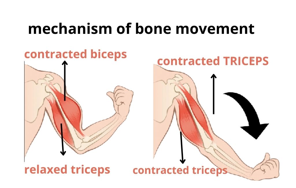
Muscular tissues are involved in the movement. They are divided into three types: skeletal, cardiac, and smooth muscle. Skeletal muscle anchors tendons to bones, while cardiac muscle pumps blood in the heart, and smooth muscle helps move food through the digestive tract and other organs. Skeletal and cardiac muscles are striated, while the smooth muscle is not.
Examples of Muscular Tissue: Tissues in biceps, heart, and intestines.
Nervous Tissue
Nervous tissue is found in the brain, spinal cord, and peripheral nerves, and is made up of neurons and neuroglia. The four types of nervous tissue are gray matter, white matter, nerves, and ganglia. Nervous tissue disorders include Alzheimer’s disease, ALS, multiple sclerosis, Huntington’s disease, and Parkinson’s disease.
Examples of Nervous tissues: Tissues in brain, spinal cord, and nerves.
Epithelial Tissue
Epithelial tissue covers the surfaces of organs, creating a protective barrier and aiding in the absorption of water and nutrients, waste removal, and enzyme or hormone secretion. All glands in the body are formed from epithelium. Epithelial tissue diseases include skin diseases like eczema and psoriasis, as well as cancers called carcinomas.
Examples of Epithelial Tissue: Tissues in skin, trachea, reproductive tract, and digestive tract.
Types Of Epithelial Tissues
Most classification schemes describe epithelium based on the shape of cells in the upper layer and the number of layers present. The two main categories are simple (one layer of cells) and stratified (multiple layers of cells). However, other cellular features, such as cilia, can also be included in the classification system.
- Simple ciliated (pseudostratified) columnar epithelium
- Simple columnar epithelium
- Simple cuboidal epithelium
- Simple glandular columnar epithelium
- Simple squamous (pavement) epithelium
- Stratified keratinized epithelium
- Stratified non-keratinized squamous epithelium
- Stratified transitional epithelium
Types of Plant Tissues
Plants are complex organisms made up of various types of tissues that work together to perform essential functions such as photosynthesis, transport of water and nutrients, and structural support.
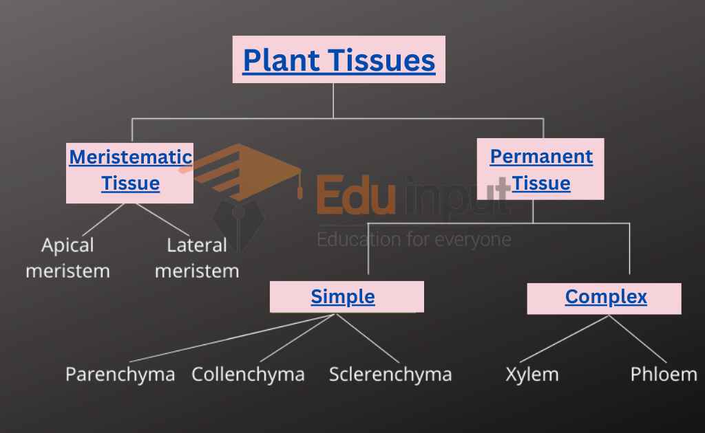
There are following types of Tissues in plants:
Vascular Tissue
Vascular tissues in plants transport substances throughout the different parts of the plant. The two types of vascular tissue are xylem and phloem. Xylem transports water and some soluble nutrients, while phloem transports organic compounds the plant uses as food, particularly sucrose.
Examples of vascular tissue are the veins in a leaf (Xylem, Phloem), which transport water and nutrients to the cells and carry away the products of photosynthesis.
Ground Tissue
Ground tissue is made up of all cells that are not vascular or dermal. There are three types of ground tissue: parenchyma, collenchyma, and sclerenchyma. Parenchyma cells form the “filler” tissue in plants and perform many functions like photosynthesis, storage of starch, fats, oils, proteins, and water, and repairing damaged tissue.
Examples of parenchyma cells are the cells found in fruits, which store nutrients for the developing seed. Collenchyma tissue is made up of long cells with irregularly thick walls that provide structural support to the plant. Plants that grow in windy areas have thicker walls of collenchyma tissue.
Sclerenchyma is also supporting tissue, but it is made of dead cells. Examples of sclerenchyma tissue are the hard shells of nuts, which protect the developing seed.
Epidermal Tissue
The epidermis is made up of a single layer of cells that covers a plant’s roots, stems, leaves, and flowers. It guards the plant against water loss, regulates the exchange of carbon dioxide and oxygen, and in roots, it absorbs water and nutrients from the soil.
Examples of epidermal tissue are the hairs on a tomato plant, which help to reduce water loss from the leaves, and the scales on a bulb, which protect the developing plant from damage.
Meristematic Tissue
Meristematic tissue consists of actively dividing cells and leads to an increase in length and thickness of the plant. The primary growth of a plant occurs only in certain specific regions, such as in the tips of stems or roots. It is in these regions that meristematic tissue is present. Cells of this type of tissue are roughly spherical or polyhedral to rectangular in shape, with thin cell walls.
Types of Meristematic Tissue
- Apical Meristem – Responsible for the linear growth of an organ, located at the growing tips of stems and roots.
- Lateral Meristem – Causes the organ to increase in diameter and girth, usually occurring beneath the bark of the tree as cork cambium and in vascular bundles of dicotyledons as vascular cambium.
- Intercalary Meristem – Located between permanent tissues, responsible for growth in length of the plant and increasing the size of the internode.
Permanent Tissue
Permanent tissues may be defined as a group of living or dead cells formed by meristematic tissue and have lost their ability to divide and have permanently placed at fixed positions in the plant body.
Meristematic tissues that take up a specific role lose the ability to divide. This process of taking up a permanent shape, size, and function is called cellular differentiation. Cells of meristematic tissue differentiate to form different types of permanent tissues.
Types of Permanent Tissue
- Simple Permanent Tissue – A group of cells that are similar in origin, structure, and function.
- Complex Permanent Tissue – Composed of more than one type of cells that work together to form a specific function.
Types of Simple Permanent Tissue
- Parenchyma – Consists of relatively unspecialized living cells with thin cell walls that are usually loosely packed so that intercellular spaces are found between cells of this tissue.
- Collenchyma – A living tissue of primary body like Parenchyma. Cells are thin-walled but possess thickening of cellulose, water and pectin substances (pectocellulose) at the corners where a number of cells join.
- Sclerenchyma – Consists of thick-walled, dead cells and protoplasm is negligible. These cells have hard and extremely thick secondary walls due to uniform distribution and high secretion of lignin and have a function of providing mechanical support.
Latest Research In Filed Of Histology
- Studies by Murphy et al. (2014), Skardal et al. (2017), and Ozbolat and Hospodiuk (2016) showcase the potential of 3D bioprinting in creating functional tissues and organs for transplantation and therapies. The studies highlight the importance of vascularization in creating tissue constructs that can mimic complex physiological microenvironments. The authors suggest optimizing bioprinting techniques and using sacrificial materials and co-culture techniques to overcome challenges in vascularization. [2]
- Research has shown that the development of an organism relies on the genetic programs of progenitor cells and their complex environment. Synthetic biology can manipulate cell communication, adhesion, and fate to improve tissue and organoid models and design tissues for regenerative purposes. [3]
Leave a Reply