Leaf Cross Section Diagram and Function of Its Parts
February 18, 2024  written by Sidra Batool
 written by Sidra Batool
Leaf Cross Section Diagram
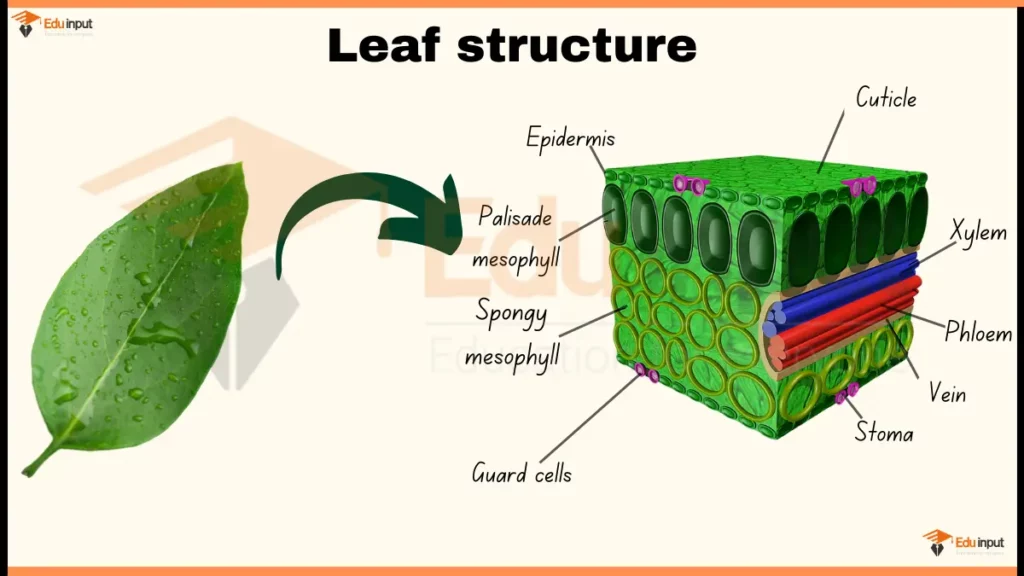
Here are the main parts of Leaf that can be observed under microscope during its cross-section:
- Epidermis: The outermost layer of the leaf, protecting it from water loss, infection, and UV radiation.
- Palisade mesophyll: A layer of tightly packed cells directly below the epidermis, containing many chloroplasts for photosynthesis.
- Spongy mesophyll: A layer of loosely packed cells below the palisade mesophyll, containing fewer chloroplasts but allowing gas exchange.
- Vein: A bundle of xylem and phloem tissues that transport water, nutrients, and sugars throughout the leaf.
- Stoma: Tiny openings in the epidermis that allow for gas exchange (carbon dioxide in, oxygen out) and some water vapor loss.
- Guard cells: Specialized cells that surround the stoma, regulating stomatal opening and closing based on environmental factors.
File Under:
Leave a Reply