Watson and Crick Model | The Double Helical Structure of DNA
 written by Sidra Batool
The structure of DNA resembles a twisted staircase and looks like a double helix. The Double Helical Structure of DNA was firstly described by Franklin.
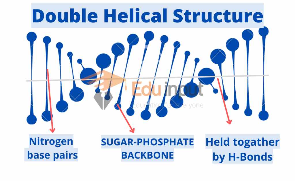
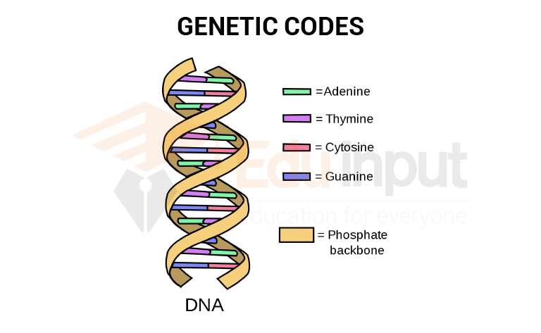
A molecule of DNA consists of two strands wound around each other as though a twisted ladder where each strand comprises a backbone comprising alternating groups of phosphate and sugar groups. The nitrogenous bases are centrally placed holding together these two strands.
One of the 4 bases is attached to each of the sugar. Both strands are united with the help of bonds between the bases. Adenine forms a base pair with thymine while cytosine pairs with guanine. This pairing is referred to as the base complementary rule since the DNA strands are complementary to one another.
The experiment of Rosalind Franklin and Maurice Wilkins (Base of this Model)
British chemist Rosalind Franklin carried out an x-ray diffraction analysis of DNA. This analysis proved the observation of the Chargeoff. He bombarded a molecule with a beam of x-rays. Rays encounter atoms.
Rosalind Franklin prepared this x-ray diffraction pattern of DNA in the laboratory of British biochemist Maurice Wilkins. Wilkins prepared DNA fibers.
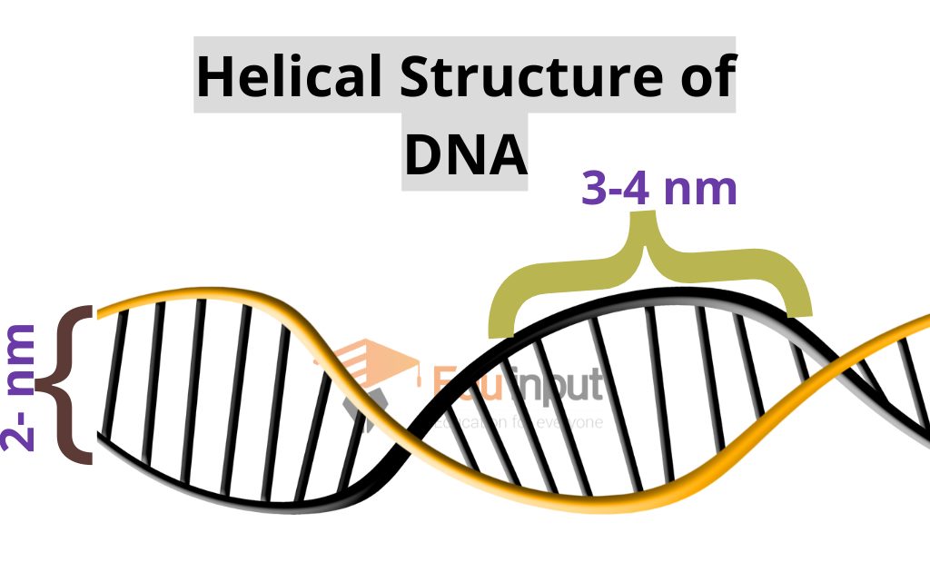
The diffraction pattern suggested that the shape DNA molecule is a helix. It has a diameter of 2m. It has a complete helical turn after every 3.4nm.
Watson and Crick Model
James Watson and Francis crick were two young researchers at the University of Cambridge England. They learned the results Franklins and they quickly worked out the structure of the DNA molecule. It is now proved that this structure is correct.
There are following important features of this model:
Double helix
The DNA molecule is a simple double helix. The basis of two strands pointed toward each other and they form base pairs.
Base pairing
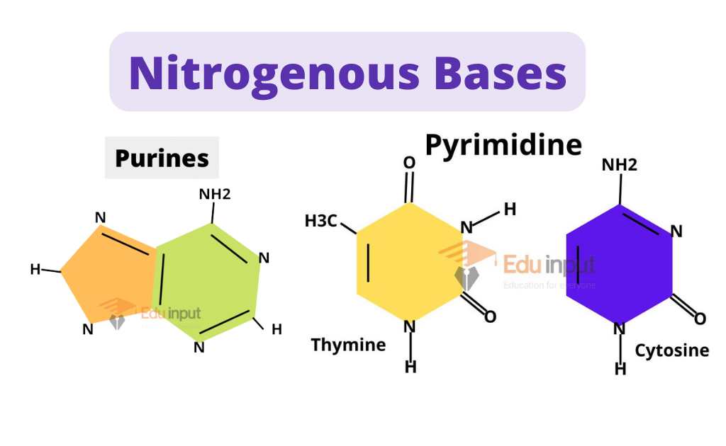
Base pairs always consist of purines and pyrimidine bases. Purines are large bases and pyrimidines are small bases. It keeps the diameter of the molecule a constant of 2 nm.
Hydrogen bonding between base pairs
Hydrogen bonds are formed between the bases in a base pair. Adenine forms two hydrogen bonds with thymine. But guanine forms three hydrogen bonds with cytosine. Adenine does not form proper hydrogen bonds with cytosine. Similarly, guanine does not form hydrogen bonds with thymine.
Thus adenine and thymine always occur in the same proportion in any DNA molecule, as the guanine and cytosine.
Antiparallel strands
The molecule of DNA is composed of two antiparallel strands. One chain runs 3’ to 5’ and the other 5′ to 3′. Thus the double helix is stabilized as a duplex DNA molecule.
Hydrophobic interactions
The base pairs are planar (flat). They stack 0.34n apart due to hydrophobic interactions. It increases the stability of the molecule.
Related FAQs
What is Watson and Crick’s model of DNA?
This model states that the structure of DNA resembles a twisted staircase and looks like a double helix. These two strands of DNA are held together by hydrogen bonding between Nitrogenous bases and form a helix.
What are Antiparallel strands?
The molecule of DNA is composed of two antiparallel strands. One chain runs 3’ to 5’ and the other 5′ to 3′. Thus the double helix is stabilized as a duplex DNA molecule.
Who discovered double helical structure of DNA?
James Watson and Francis Crick discovered double helical structure of DNA.
Which technique was used to discover Helical structure of DNA?
James Watson and Francis Crick used X-ray crystallography to see the structure of DNA.
Leave a Reply