Connective Tissues-Definition, Structure, Types, Functions, and Examples
 written by Sidra Batool
Connective tissue is an essential tissue that connects, separates, and provides support to all other tissues in the body. It is made up of cells surrounded by a fluid compartment called the extracellular matrix (ECM). Unlike other types of tissue, connective tissue has loosely packed cells within the ECM.
What is Connective Tissue?
Connective tissues are tissues that are composed of ectracellular matrix and limited number of cells scattered in ectracellular matrix . They helps our body to maintain the shape of our organs and provides support to our body.
It is made up of several types of fibrous tissues. These tissues differ in their density and cellular structure. Connective tissue also includes some specialized tissues like bones, ligaments, tendons, cartilage, and fat tissues.
Location Of Connective Tissues
Connective tissues are distributed throughout the body. They are found in the meninges that wrap the brain and spinal cord, loose connective tissue beneath the skin, and dense irregular connective tissue in the dermis.
Structure of Connective Tissues
Connective tissue has three main components:
- Extracellular Fibers
- Stationary And Migrating Cells
- Ground Substance
The amount of these components in connective tissue can vary depending on the specific function of the tissue in different parts of the body.
The mechanical and biological properties of connective tissues are intricately tied to their chemical composition, which is made up of a complex system of interacting macromolecules.
These properties can be understood by applying concepts developed to analyze the behavior of fiber-reinforced composite materials. [1]
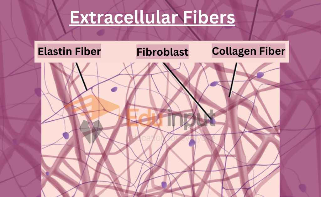
1: Extracellular Fibers
Loose and dense connective tissue are two types of connective tissue that are composed of collagen fibers, reticular fibers, and elastin fibers.
Collagen Fibers
Collagen fibers are tightly packed thin fibrils that offer flexible but powerful resistance to pulling force. They run in a parallel course in loose connective tissue, creating a three-dimensional meshwork.
In dense connective tissue, such as ligaments and tendons, collagen fibers are densely packed.
Reticular Fibers
Reticular fibers are fine, dark fibrils that form a network underlying the basal lamina layer. They are present in basement epithelial tissue, adipose cells, Schwann and muscle cells, lymphoid tissue, and endothelium of hepatic sinusoids.
These fibers are continuous with collagen fibers and have a firm attachment to the basal lamina.
Elastin Fibers
Elastin fibers have the property of elastic recoil and are present in a loose network in loose connective tissue. They are present in a concentric form in the vascular wall to help maintain uniform blood pressure.
These fibers are also present in distensible and contractible organs such as the lungs and urinary bladder.
2: Ground Substance
Connective tissues constitutes following ground tissues:
- Large carbohydrate molecules or complexes of protein and carbohydrate called glycosaminoglycans (formerly known as mucopolysaccharides)
- Hyaluronic acid, composed of glucuronic acid and an amino sugar, N-acetyl glucosamine
- Chondroitin-4-sulfate (chondroitin sulfate A) and chondroitin-6-sulfate (chondroitin sulfate C), composed of galactosamine and glucuronate sugars
- Multiple chains of chondroitin sulfate bound to protein
- Viscous solution or highly hydrated thin gel consistency
3: Stationary And Migrating Cells
Fibroblasts are the main stationary and active cells found in connective tissue. They have a long, spindle-shaped appearance and are located along bundles of collagen fibrils. Fibroblasts secrete tropocollagen and other components of the ground substance to maintain the extracellular tissue components.
Alongside the fixed cell types (stationary cells) found in connective tissue, there are also free cells that can move through the extracellular spaces. One of these types is mast cells, which are characterized by their cell body containing granules filled with histamine and heparin.
These granules are biologically active and can be released in response to mechanical or chemical irritation. When Histamine is released, it causes fluid to escape from nearby blood vessels, that results in local swelling. This is commonly seen in the form of a welt around an insect bite. While, Heparin prevent or delay blood clotting when added to the blood.
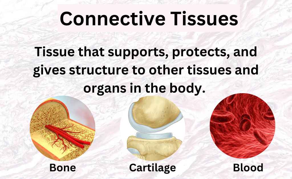
Types Of Connective Tissues
Types of connective tissues include loose connective tissue, dense irregular connective tissue, adipose tissue, cartilage, bone, blood, and lymph.
There are two main types of connective tissue based on cellular and ECM characteristics.
1: Connective Tissue Proper
They can be further subdivided into loose and dense connective tissues.
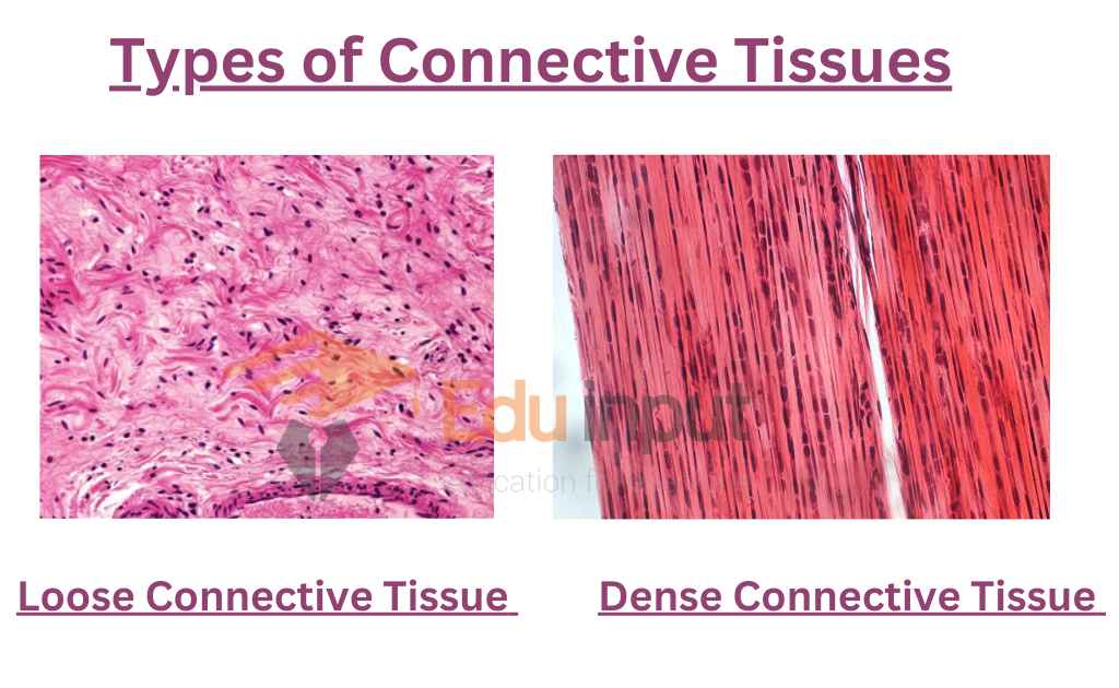
Loose Connective Tissues
Loose connective tissue, also known as areolar connective tissue, is composed of nearly equal amounts of cells, fibers, and ground substance. The primary cell type is the fibroblast, but immune system cells are also present.
Collagen fibers are the most abundant type of fiber in the extracellular matrix (ECM) but are sparsely distributed, hence the name “loose.” Other types of fibers, such as reticular and elastic fibers, are also present in moderate amounts.
Loose connective tissues are widely distributed throughout the body, lining the inner surfaces of organs. The combination of cells and fibers provides the tissue with flexibility but not high resistance to mechanical stress.
Loose connective tissue plays an important role in binding different types of tissues together, joining them into organs, holding organs in place, and attaching epithelial tissue to other tissue types. The presence of immune system cells also gives it an important immuno-protective function.
Functions Of Loose Connective Tissues
- Provides support and elasticity where needed throughout the body
- Wraps around blood vessels, nerves, and muscles
- Acts as a shock absorber and reservoir for salt and fluid
- Contains areolar tissue and adipose tissue, which support organs, fill spaces, and store fat
- White adipose tissue protects organs and helps maintain body temperature, while brown adipose tissue generates heat
- Reticular connective tissue supports the internal framework of organs such as the liver, lymph nodes, and spleen
2: Dense Connective Tissue
In comparison to loose connective tissue, dense connective tissue has a lower number of cells and a higher density of collagen fibers in its ECM. There are two subtypes of dense connective tissue, namely dense regular and dense irregular, distinguished by the arrangement of the fibers.
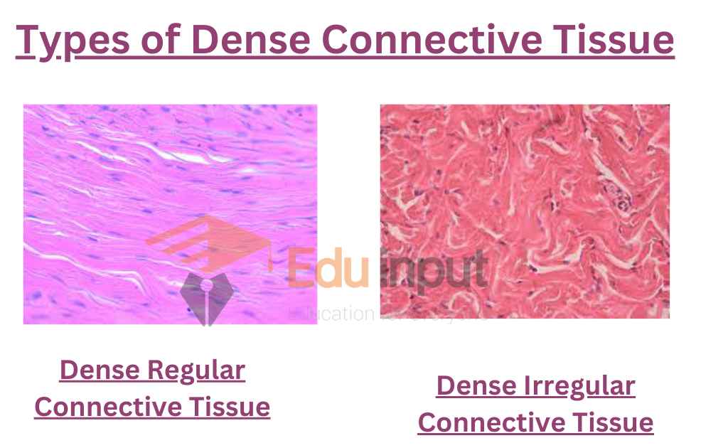
Dense Regular Connective Tissue
Dense regular connective tissue consists of parallel-aligned collagen fibers, which provide high unidirectional resistance to stress. Tendons and ligaments are examples of dense regular connective tissue.
Dense Irregular Connective Tissue
In contrast, dense irregular connective tissue consists of collagen fibers that interweave randomly. They create a three-dimensional network resistant to distension in all directions. This type of tissue is commonly located in organ capsules and walls, the dermis of the skin, and glands.
Functions Of Dense Connective Tissue
- Its Main function is to support and transmit mechanical forces
- Dense regular tissue, with regular orientation of fibers, enhances tensile strength and resistance to stretching in the direction of fiber orientation
- Dense irregular tissue, with irregular or random orientation of fibers, provides uniform strength in all directions and forms a mesh-like network
3: Specialized Connective Tissue
It includes reticular, blood, bone, cartilage, and adipose tissues.
Cartilage
Cartilage Cartilage is mostly present in the embryonic stages and works as a supporting skeleton. Most of the cartilage is replaced by bones in adults, however, it supports some structures in adults too. In humans, cartilage is present between the bones of the vertebral column, in the external ear, nose and hands.
The cartilage consists of chondrocytes cells, which are enclosed in a hard, rubbery matrix, secreted by them. They secrete collagen fibers also, which provide additional strength. Chondrocytes lie in the cavities known as lacunae, in a group of 2-4 cells or singly.
Bones
Bone is the hardest connective tissue and helps in maintaining the shape and posture of the body, it protects internal organs. They are rich in collagen fibres and calcium, which give strength.
The cells of the bone are known as osteocytes. They are present in lacunae and secrete the matrix. There is substantial blood supply in bony tissues. The cytoplasmic extension of osteocytes makes tiny channels known as canaliculi. These channels help in communication among osteocytes and capillaries.
Blood and Lymph
Blood is a fluid connective tissue composed of various cells suspended in a liquid matrix called plasma. The cells in blood include Red blood cells (RBCs), White blood cells (WBCs), and platelets. RBCs contain the protein hemoglobin and are responsible for transporting oxygen to tissues throughout the body. WBCs form part of the immune system, defending the body against foreign invaders such as bacteria and viruses. Platelets play an essential role in blood clotting to prevent excessive bleeding.
Plasma contains a variety of substances, including proteins, water, hormones, and salts, which transport to different parts of the body.
Lymph, another fluid connective tissue, drains into the blood and transports absorbed fat to the bloodstream, which cannot enter directly. Lymph contains WBCs that help in removing toxins and waste materials from the body and fighting infection.
Examples Of Connective Tissues
Here are some examples of connective tissues:
- Loose Connective Tissue (Areolar Tissue)
- Dense Irregular Connective Tissue
- Adipose Tissue (Fat)
- Cartilage
- Bone (Osseous Tissue)
- Blood
- Lymph
- Dense Regular Connective Tissue (Found in tendons and ligaments, among other places)
- Reticular Tissue (Found in organs like the spleen and lymph nodes)
10 Functions Of Connective Tissues
Most important functions of connective tissues are:
- They provide support and structure to the body
- Binds and connects different tissues and organs
- Protects and cushions internal organs
- Acts as a storage site for fat
- Plays a role in immune defense
- Loose connective tissue: binds tissues together, provides flexibility and support, and contains immune system cells
- Cartilage supports structures in the body, is elastic and firm, and lacks nerves, blood vessels, and lymph vessels
- Bone maintains shape and posture of the body, protects internal organs, and contains osteocytes that secrete matrix and help with communication
- Blood contains red blood cells that transport oxygen, white blood cells that protect from foreign antigens, platelets that aid in blood clotting, and plasma that transports proteins, water, hormones, and salts throughout the body
- Lymph drains into the blood and helps transport absorbed fat, contains white blood cells that fight infections and rid the body of toxins and waste materials.
Latest Research In Field Of Connective Tissues
- Recent studies have shown that a proper gingival connective tissue thickness is essential for dental implant and reconstructive surgery success. This study compared the clinical outcomes of gingival augmentation using palatal connective tissue grafts and a xenogenic soft-tissue substitute. Both methods showed better results in maintaining the alveolar bone level and thickness of the attached gingiva compared to the control group with no augmentation. The soft-tissue augmentation with connective tissue grafts before implant placement was found to be the most efficient method, providing a stable increase in tissue thickness. [2]
- A recent 5-year follow-up study evaluated soft-tissue augmentation around dental implants using a connective tissue graft (CTG) and a xenogeneic collagen matrix (CMX). The study found both methods to have better clinical outcomes in maintaining the alveolar bone level and thickness of the attached gingiva compared to the control group. CTG before implant placement was found to be the most efficient method for a stable increase in tissue thickness. [3]
- Recent research has shown that there is a functional relationship between the buccinator muscle and connective tissue in the cheek. Fibrous septa containing collagens and elastic fibers pass through the muscle from the submucosa to the skin, indicating the potential for the connective tissue to affect the muscle’s function. Further study is needed to fully understand this relationship. [4]
FAQs
Where is connective tissue found?
Connective tissue is found everywhere in the body between other tissues, offering support, structure, and connect.

Leave a Reply