Skeletal Muscle Fiber-Definition and Structure
 written by Sidra Batool
Each muscle consists of muscle bundles. Each muscle bundle is further composed of skeletal muscle fiber or muscle cells. Each muscle fiber is a long cylindrical cell the diameter of the muscle fiber cell is 10-100 micrometers. It has the following structure;
Nuclei
Muscle fiber cells contain multiply oval nuclei. These nuclei are arranged just beneath the sarcolemma.
Sarcolemma
The outer membrane of the muscle cell is called sarcolemma. But it contains a large amount of stored glycogen and myoglobin. Myoglobin is a unique oxygen-binding protein. It has a red pigment that stores oxygen.
Myofibrils
Each muscle fiber contains a large number of myofibrils, 1-2 micrometer in diameter. These fibers run in a parallel direction to the muscle fiber they extend the entire length of the cell. Sarcolemma encloses bundles of myofibrils.
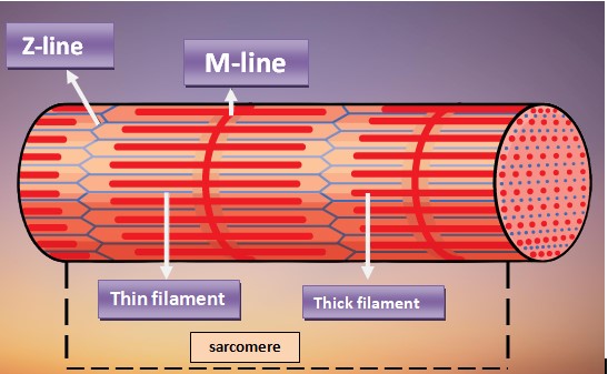
Sarcomere
The small contractile unit of the myofibrils is called a sarcomere. A sarcomere is the region of myofibrils between two successive Z-lines. The myofibril contains myofilament. Each sarcomere has a series of dark and light bands. These bands can be seen along the length of each myofibril. There are the following parts of a sarcomere.
A-band
The dark bands are anisotropic so they are called A bands. An anisotropic compound can polarize visible light.
I-band
The light when called I band. They are the isotropic or nonpolarizing band. These give the cell are striped appearance.
H-zone
Each A-band has a lighter strip in its mid section. This lighter strip is called the H zone. H stands for Hele, it means bright.
M-line
The H zone is bisected by Dark Line called the M line.
Z-line
The I band has a midline called Z- the line. Z stands for zwish means between.
Structure of Myofilament
Myofilaments are composed of thick and thin filaments;
Thick filament (Myosin)
The central thick filament extends to the entire length of the band. A thick filament is about 16 nm in diameter. It is composed of Myosin protein. Each Myosin molecule has a tail. The tail ends in two globular heads.
Myosin tail consists of 2 long polypeptide chains coiled together. The heads link the thick and thin filament together during a contraction so they are sometimes called cross bridges.
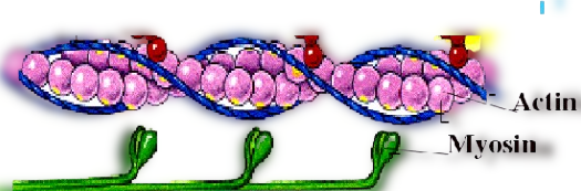
Thin Filament
The filament is 7 to 8 nanometers. it is composed of 3 proteins.
Actin
Thin filaments are composed of the chiefly acting molecule. The thin filament extends across the I-band and partially into the A band. The actin molecule is arranged in two chains. These chains twist around to surround each other like twisted double strands of pearls.
Tropomyosin
Two strands of another protein tropomyosin also twist around the acting.
Troponin
The other major protein in the thin filament is troponin. It is three polypeptide complexes. One binds to actin, another binds to tropomyosin, and the third binds calcium ions. Myosin filament is surrounded by 6 actin filaments on each end.
Frequently Asked Questions-FAQs
When does a skeletal muscle fiber contract?
As cross-bridges are formed between the filaments of actin and myosin proteins and shortens the sarcomere, which causes contraction of skeletal muscle fibers.
Which layer of connective tissue surrounds each layer of skeletal muscle?
Endomysium surrounds each layer of skeletal muscle.
How many nuclei are present in skeletal muscle fiber?
The number of nuclei depends upon the fiber’s length and size. So a skeletal muscle can be multinucleated. It ranges from 50-to 1000 per fiber.
What is the Functional unit of skeletal muscle fiber?
The sarcomere is the functional unit of skeletal muscle fiber.
What are the major parts of skeletal muscle fibers?
There are two major components of skeletal muscle fiber;
i) Thin filament (containing actin, troponin, and tropomyosin).
ii) Thick filament (containing actin).
What is the repetitive unit in skeletal muscle fiber?
In a skeletal muscle fiber, a sarcomere is a repetitive unit between z-lines.
Where is ATP consumed in skeletal muscle fibers?
Hydrolyzation of ATP occurs when the sliding of actin and myosin starts. This energy is provided by mitochondria present in the muscle cell.
What causes relaxation in skeletal muscle fiber?
When the signal of the nervous system is no longer present, skeletal muscle fibers transport calcium back into the reticulum of sarcoplasm by using ATP. And when troponin moves back to the position to block the binding site of myosin and actin.
Leave a Reply